Know your spinal cord – Tract organization
For those of us just tuning in, today is the third part in a … well a lot of posts on the spinal cord! If you’re just joining us, you should probably start from the top (literally) here and this post covers the anatomy of the cord. Today we are going to talk spinal organization, for that reason we also should talk about how the brain is organized, which will help us make sense of why the spinal cord is organized the way it is, so let’s get started!
The motor cortex, it is where we start deliberate movement. Like the spinal cord, the brain has a very specific organization to it. We normally draw this out using the homunculus, which represents the area of the brain associated with the body part. Behind the motor cortex is the somatosensory cortex, this handles all afferent information (which is sensory information, remember Afferent signals Arrive at the brain). It too has an organization and in the image below, we see where on the brain the two areas are and how they are broken up.
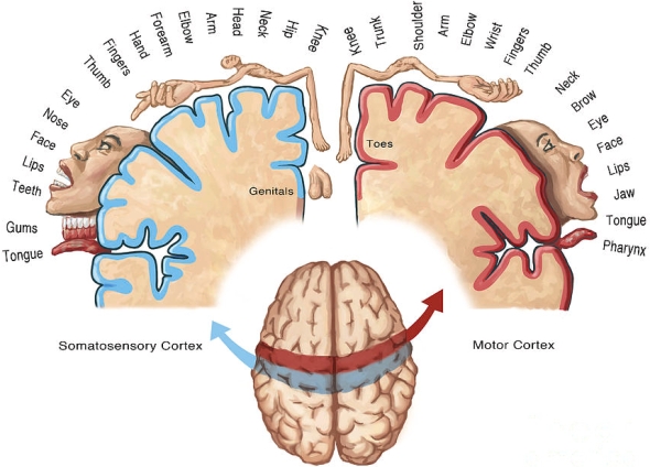
The motor cortex (red strip) has a similar organization to the somatosensory cortex (blue strip).
So from an EEG recording standpoint (for more info on EEG and recording from the brain read this), because we have trouble localizing the signal recording leg or foot movements proves to be difficult becuase we cannot easily tell left foot from right foot since they are so close together. The image above only shows half of each cortex areas, but it is mirrored for the other side, so we have left half of the body on the right side, and right side of the body on the left. However, we have a very easy time telling left hand from right hand because they are physically separated in two distinct areas of the brain.
Why are we talking about brain organization when we’re interested in the spinal cord? That is because the spinal cord has its own very distinct organization. Nerve axons run the length of the cord in bundles, we call these bundles fasciculi. These bundles make up different tracts of the spinal cord and we will cover them individually later, but today let’s introduce them briefly. So let’s look at a cross section of the spinal cord. In the image below we can see each of the tracts individually labeled.
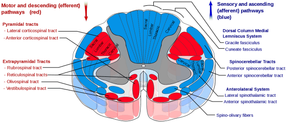
The first thing to notice, we have efferent and afferent pathways located in distinct groups. Now remember, the spinal cord like the brain is a mirror image of itself, so while only half of the tracts are labeled, the other side has the same tracts for that half of the body (also recall the left side of the cord controls the left side of the body, unlike the brain). So with that out of the way, let’s talk about what each of the tracts do in the rough sense.
First up, the dorsal medial lemniscus. This is broken into the fasciculus gracilis and cuneatus. The gracile fasciculus carries sensory information from the lower half of the body from nerves entering the spinal cord at the lumbar level. While the cuneate fasciculus carries sensory information from the upper half of the body from nerves entering the spinal cord at the cervical level. These guys are responsible for fine touch and proprioception. Proprioception is how you tell where your arm is in space. If you close your eyes and extend your arm, you know it is extended, this is proprioception and is really just the way the body measures stretch of the muscle. It’s actually a very complex calculation, but hey it manages to do it without you thinking about it.
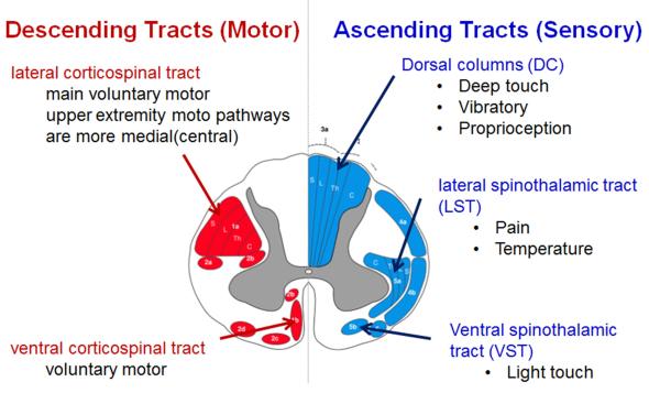
Next, we can talk about the lateral corticospinal tract. This is the descending (efferent) pathway and connects the motor cortex to the spinal cord (hence corticospinal). This guy is responsible for limb movement. In the image above, only one side is labeled, remember it is mirrored, so the same red area on the right side of the image is the same pathway for that side of the body. You’ll also notice there is an anterior corticospinal tract. This tract is responsible for the trunk muscles, which is (probably) why it is much smaller and has its own unique things going on which we will talk about. Below is an image with the main tracts, more clearly labeled. I like the one above because it shows the complexity of the cord, while the one below helps simplify things.
We also have the lateral spinothalamic tract, which is responsible for pain and temperature sensing. Like the corticospinal tract, there is a second ventral spinothalamic tract which covers light (or crude) touch. We will go over the difference between the difference between crude touch and deep (or fine) touch when we talk about the two tracts in seperate posts.
You’ll notice that the grey matter (that butterfly shape in the middle if you missed the other posts), doesn’t have anything labeled. Remember that this is because the grey matter handles processing not transmission. Don’t worry though, we will get into the grey matter as well. You’ll see that it is a very important part of the spinal cord and is what makes the spinal cord more than just a freeway for neural traffic. In fact, the amount of grey matter to white matter depends on where you are looking at the cord. In fact, let’s take a moment to show this. In the image below we can see cross sections of the spinal cord at different levels. You’ll notice the shape and amount of grey matter change as you travel along the cord. There is a good reason for this and we will discuss this soon.
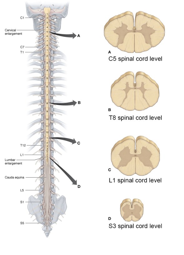
Notice at the T8 spinal cord level, there is very little grey matter, this suggests there isn’t a lot of processing going on here. We somewhat expect this because the cervical and lumbar enlargements are where the upper and lower limbs innervate at.
Tomorrow we can dive into the corticospinal pathway. This is the efferent (or descending) pathway responsible for movement! Later we will find out why 10% of the nerves do not decussate at the medullary pyramids when we get into the afferent (or ascending) pathways. Spoiler, they still decussate, but it gets really… well weird. So stick around!


But enough about us, what about you?