Know your spinal cord – The anatomy
Now that we took it from the top, let’s get an overview of what exactly makes up the spinal cord. There is a lot, so we’re not going to do a comprehensive review since that would be a whole class and not a single post. Most of the structures we cover today, will have a seperate post where we can go into detail.
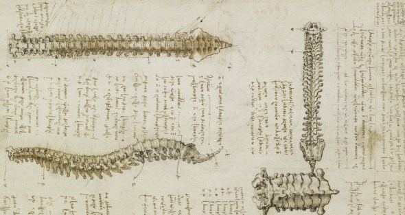
Yesterday we introduced the medullary pyramids. So we can go directly below the pyramids and start talking cord. The spinal cord is very thin, between 1 to 1.5 cm (or roughly 0.3 to 0.5 inches for those of us who are stuck on imperial units) in diameter. That makes it roughly the size of your pinky finger. Like the brain, it is made up of the same grey and white matter. White matter transmits data, grey matter processes data (in our current understanding of neurons). The brain has a grey matter exterior and a white matter interior. Interestingly enough, this is reversed for the spinal cord, the grey matter is on the inside as seen in the cross section of the cord below.
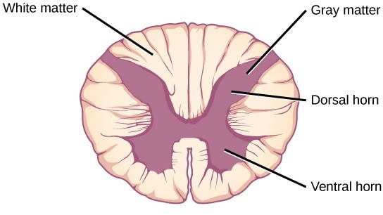
The cord is roughly symmetrical as you can see, the grey matter interior forms a sort of butterfly shape and we have the dorsal and ventral horns labeled. Dorsal is another fancy term meaning in the back and ventral means in the front, so from that we can tell which side is closest to the back and which is furthest away, which is why we use those fancy terms.
You’ll notice I gave a range for the diameter of the spinal cord, well that’s for two reasons, one is variability in the individual, but the other is part of the anatomy. There are two enlargements of the spinal cord, the cervical enlargement and the ventral enlargement. The cervical enlargement is located at the back of the neck (cervical giving us the location) and the lumbar is located in the lower back (again lumbar is the location). We can see these enlargements in the image below. The cervical enlargement controls the arms (primarily) and the lumbar enlargement handles the legs (again, primarily).
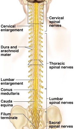
An interesting fact, the spinal cord doesn’t run all the way down the back, it tapers off and comes to almost a point. We call that the conus medullaris (seen labeled above). The rest of the spinal column has a bunch of nerve fibers that are called the cauda equina. Equina might sound familiar, the cauda equina gets its name because it looks like a horses tail. To keep the cord from flopping around when you move it is held in place by a long fiber called the filum terminale which translates roughly to the terminal thread, which sounds more ominous than it is.
The cord shares a lot of other interesting similarities with the brain, it is covered in the same protective membranes (called the meninges) and even shares cerebrospinal fluid with the brain. We can go into detail about the protective layers and what they do another post, but to give you an overview of the coverings, we have a good example of it below.
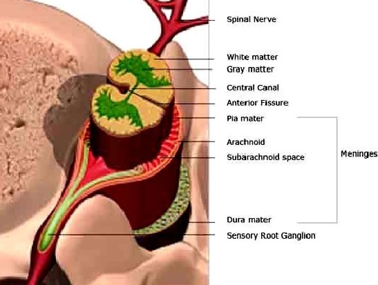
This leads us to the fibers coming off the cord! These guys are what send and receive information. There are two sets of roots coming off each level of the spinal cord running the full length from cervical all the way to lumbar and each innervate (handle) a different part of the body. The two sets coming off the cord are afferent and efferent pathways. The way I remember this one, Efferent signals Exit the brain, while Afferent signal Arrive at the brain. Maybe it’s stupid, but it works for me and chances are it will work for you too. You can see a better view of the rootlets (the nerves coming off the cord) below.
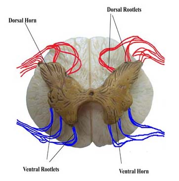
The rootlets marked in red are the afferent pathway and the ones in blue are the efferent pathway. I know this because they are labeled as dorsal and ventral, see anatomy terms come in handy! Remember, the left side of the cord controls the left side of the body, while the right side of the cord controls the right side of the body. This helps to explain why the spinal cord is symmetric. The dorsal rootlets run through something called the dorsal root ganglion, which only exists for the afferent pathway. We can see this more clearly in the image below.
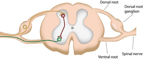
Inside the dorsal root ganglion is a pseudounipolar neuron, yet another fancy term which tells us the type of neuron that is located there. We will go into detail some other time about the dorsal root ganglion and it’s pseudounipolar neuron. The last thing we should mention is that there is a hole in the center of the spinal cord, you can see it in the drawing above. That is called the central canal and below is another image with the central canal labeled. The central canal is filled with spinal fluid, which as we mentioned before is shared with the brain.

That pretty much covers the gross anatomy of the spinal cord. Next up I think we will talk about how the spinal cord innervates the body. Like the brain, there is a clear organization to how and where the spinal cord innervates to the body! Eventually we can talk about spinal cord misconceptions too. One fun fact, the spinal cord was once thought to be the freeway of nervous system. It was only in charge of sending and receiving information, but we now know the spinal cord can learn and have “seen” it using fMRI. Now you may see why I’m so excited to talk about the spinal cord!


But enough about us, what about you?