Know your spinal cord – The landmarks
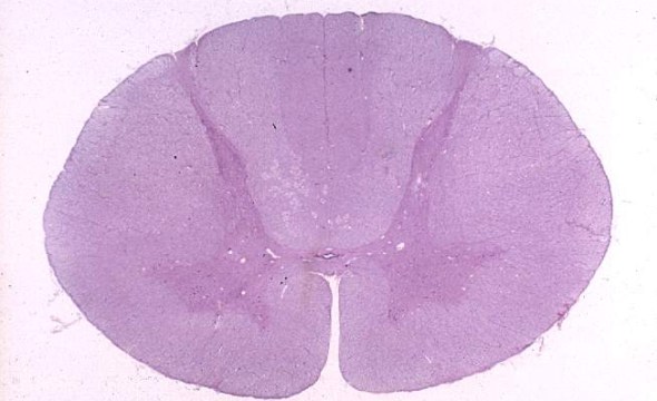
A human cervical spinal cord slice. Once you’ve read this post, you should be able to identify the major parts and tell which side is the front of the cord.
Welcome to day thirty-three in our series. For those of you who are just finding us, we have every one of these posts in our neuroanatomy category in reverse chronological order. Today we’re going to backtract (get it?) a little and go over something basic, but something we’ve skipped over to this point. We never really talked about the landmarks of a spinal cord slice. So today, we are going to take a detour and go over spinal cord features.
Chances are, if you’ve lived in one place for some time you know where you are in relation to the things around you. If I were to drop you off in the middle of some place familiar to you, you would be able to orient yourself and find your way to wherever it is you want to go. Over the course of our series I did my best to help orient us by pointing out different features of the spinal cord, but we never really defined what those were explicitly and seperate from the other things we were covering. Had I planned this series better, I may have done this post closer to the beginning. Probably after the gross anatomy of the cord, which was similar to today. However, we never really touched on the landmarks we use when we talk about the spinal cord. Well, we’re here now so let’s go over it.
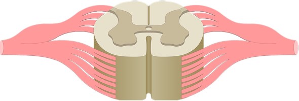
Above you see a transverse slice of spinal cord. If you’ve been following along, you know the grey matter, the white matter and you may remember a lot of the features that we’ve covered. You may even recall what we are covering today. If you’re looking at this slice of cord and are wondering which side is the anterior side and which is the dorsal, well that is why we are covering this topic today, so let’s get our frame of reference. First, let’s talk about the anterior median fissure (labeled below as ventral median fissure, ventral and anterior are interchangeable terms here, but I personally prefer ventral due to the way it’s defined. Note that there is a difference, but they are the same directions in humans), which is a very prominent spinal cord feature that will help orient you next time you see a slice like this.
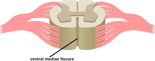
The anterior median fissure is super helpful in orienting us when we are talking about the spinal cord. Anterior (or ventral) means towards the front. So if I could see through you and look at your spinal cord, I would have to look through your chest if I wanted to see the anterior median fissure. Below is a transverse spinal slice. You can see the anterior median fissure and this landmark orients us when we’re looking at the spinal slice.

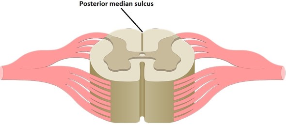
Next, we can point out the posterior (or dorsal) median sulcus. Sulcus is a fancy anatomy term meaning a long narrow slit (or a groove) that divides an organ into lobes. While the anterior median fissure is clearly two distinct and separate parts, the posterior median sulcus appears as two lobes that are joined together. Below is an image highlighting the posterior median sulcus so you can see what I am talking about.
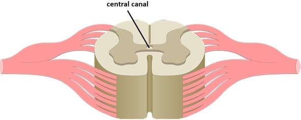
Now we have two landmarks that tell us the front of the cord and the back of the cord. We also can point out the central canal which as the name suggests, it is a canal located centrally in the spinal cord that is filled with cerebrospinal fluid. It runs the full length of the spinal cord and while this doesn’t tell us front and back, it does help us locate center. Below we have an image with the central canal labeled for you.
Because the spinal cord is a rough mirror image of itself, we cannot really determine left side from right side, just front and back. While using the anterior median fissure or posterior median sulcus let’s us do that, there are cases where you may see an image of just a portion of the spinal cord, in this case both landmarks might be out of view. In this case we have two other landmarks we can use to determine front and back.
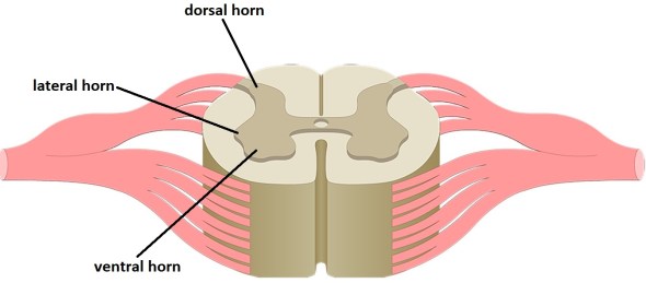
We can use the grey matter to tell us front and back of the spinal cord. To do this we can use the dorsal (posterior) horn and the ventral (anterior) horns. The dorsal horn is the more narrow of the two large horns of the spinal cord. Technically we have three, a smaller (by comparison) lateral horn. The ventral horn by comparison is more rounded and spread out. Below we have them labeled so you can see what I am talking about. As a reminder, the spinal cord is a mirror of itself, so the ventral and dorsal horns are on both sides of the cord and match the labeled counterpart.
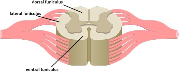
Lastly, we can define the funiculi. A funiculus is another fancy medical term meaning a bundle of nerve fibers enclosed in a sheath of connective tissue. For the spinal cord, there are three you should know, the dorsal, the lateral, and the ventral funiculi. As with most things involving the spinal cord, this applies to both sides of the cord, even though we will only be showing the labels on one side. Below we’ve labeled these and as you might imagine, the name tells you the location.
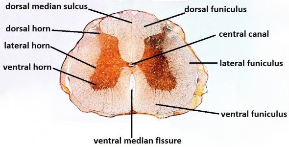
That pretty much wraps up the landmarks of the spinal cord. However, to this point we’ve only seen one image of an actual spinal cord. Here is a transverse slice of a human spinal cord that I’ve labeled with the things we’ve covered today. While the lateral horn isn’t as pronounced in this image, I’ve labeled it anyway. The image we used at the top of this post has very pronounced lateral horns (cervical spinal cross sections will have those very pronounced laterals horn), we didn’t label them because by now you should know where that is. You’ll also notice that the ventral median fissure looks like it joins together, while it is touching, they are two separate and distinct parts. The dorsal median sulcus is hard to see in this image as well, you’ll see a line in the tissue by the labeled line, this is the sulcus.
To recap, we’ve gone over the landmarks of the spinal cord. Now when we talk about where things are, we can talk about them in relation to these landmarks. Just like you might tell your friend that your house is near a particular store or cross street. This just makes it easier for us to talk about where in the spine things are located.
Now for the next post, things are busy for me this week. It’s tough to find time to write these posts when I’ve got so much going on. Never fear, they will get done, I’m just not sure what I will be covering tomorrow. Eventually our spinal series will come to an end, but once we get done talking about the anatomy, we can get into the research some. In other words, there are still some posts that will happen before we close the series and move on to something else.
Until next time, don’t stop learning!


But enough about us, what about you?