Know your spinal cord – The reticulospinal tracts

Reconstruction of the nuclear masses of the brain stem
It’s day thirty-six in our spinal cord series and I yesterday I lied, we’re not done quite yet. First, as always we have a super helpful neuroanatomy category for anyone wanting to read the posts from this series. For the rest of us, today we’re talking about the reticulospinal tracts, yes tracts with an s. There is a good reason for this, but you’ll have to read on to see why.
While it is true, we’re nearing the end of our spinal cord series, there are still things to learn. Originally I wasn’t sure if I wanted to cover the reticulospinal tracts, once we get into it you’ll see why. However, it makes sense because this could potentially come up in conversation (specifically if you work with the spinal cord like I do). So let’s go over this a little bit.
The reticulospinal tracts are what we call a group of tracts. This means it isn’t one tract specifically, but a set of tracts that have a certain function. We’ve seen in previous posts that there are multiple tracts that handle the same type of information or perhaps complementary information. The reticulospinal tracts are what we call a particular group of these complementary tracts.
First let’s go over the overall function of the reticulospinal tracts, then we can break it down into what tracts specifically are grouped here. As usual, the name gives us some information about these tracts, they are the reticulospinal tracts, meaning they end in the spinal cord and start at the reticular formation of the brain (technically brain stem and seen below). Therefore without knowing anything else, we know where the tracts originate and where they terminate. This also tells us that the tracts in this group are descending or efferent tracts.
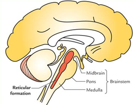
Now let’s talk about the function, these tracts are extrapyramidal motor tracts (meaning they don’t go through the medullary pyramids) that descend from the reticular formation (shown above) in two groups of tracts to act on the motor neurons supplying the trunk and proximal limb flexors and extensors. This tells us that the reticulospinal tracts are involved mainly in locomotion and postural control, although they do have other functions as well.
The descending reticulospinal tracts are just one of four major cortical pathways to the spinal cord for musculoskeletal control. The reticulospinal tracts work with the other three pathways to give a coordinated control of movement. The four pathways can be grouped into two main system pathways – a medial system and a lateral system. The medial system includes the reticulospinal pathway and the vestibulospinal pathway, and this system provides control of posture. The corticospinal and the rubrospinal tract pathways belong to the lateral system which provides fine control of movement. The reticulospinal tracts are two distinct tracts, the medial and lateral reticulospinal tracts. Below is an image showing all of the extrapyramidal tracts, the second tract listed is the reticulospinal tracts and shows the location for both the medial and lateral tracts. The only tract listed here that we haven’t explicitly mentioned (for good reason) is the olivospinal tract.
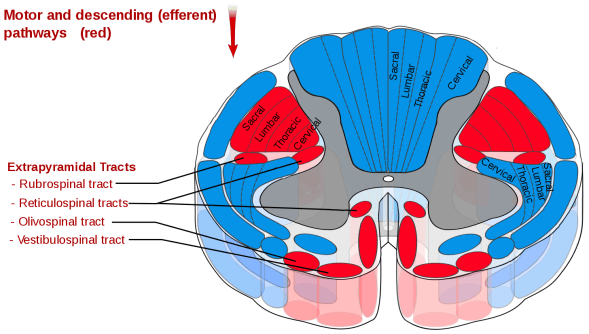
Location of the extrapyramidal tracts of the spinal cord
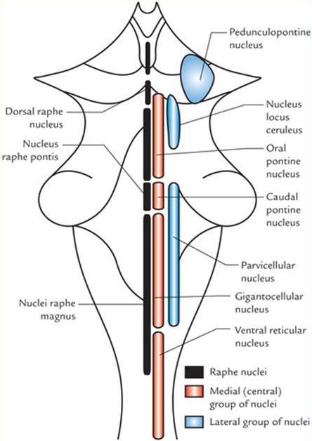
The two tracts are separated because they have two different functions. The medial tract is responsible for exciting anti-gravity, extensor muscles (which we first mentioned in our vestibulospinal tract post). The fibers of this tract arise from the caudal pontine reticular nucleus and the oral pontine reticular nucleus and project to lamina VII and lamina VIII of the spinal cord. For reference, the nuclei of the brain stem can be seen below.
The lateral tract is responsible for inhibiting excitatory axial extensor muscles of movement. It is also responsible for automatic breathing. The fibers of this tract arise from the medullary reticular formation, mostly from the gigantocellular nucleus (also seen below), and descend the length of the spinal cord in the anterior part of the lateral column. The tract terminates in lamina VII mostly with some fibers terminating in lamina IX of the spinal cord.
So there you have it, the reticulospinal tracts! A slightly complicated topic, but one I think needed to be covered. Now let’s talk about tomorrow’s post… well if there are reticulospinal tracts, could there be spinoreticular tracts as well? It’s a safe bet and tomorrow we can talk about what those do (hint: it’s mostly the same … mostly).
Until next time, don’t stop learning!


But enough about us, what about you?