Day 326: Review: The state of spinal cord research
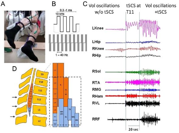
Facilitation of stepping-like volitional oscillations using non-invasive transcutaneous electrical spinal cord stimulation in SCI subject. (A) Position of the participant in the gravity-neutral apparatus. (B) Biphasic electrical stimulation was delivered using unique waveforms consisting of 0.3–1.0 ms bursts filled by 10 kHz frequency that were administered at 5–40 Hz. (C) EMG activity of right soleus (RSol), right tibialis anterior (RTA), right medial gastrocnemius (RMG), right hamstrings (RHam), right vastus lateralis (RVL), right rectus femoris (RRF) and angular displacement in the knee and hip joints of both legs during leg oscillations with a voluntary effort alone (Vol), stimulation at T11 (Stim), and Vol + Stim are shown. (D) Schematics demonstrating the approximate location of transcutaneous electrodes above the lumbosacral enlargement, in relation to the location of the motor pools based on Kendall et al. (1993) and Sharrard (1964).
Well it’s been two weeks (roughly) and my PI asked specifically that this week I do a review on the state of spinal cord research, with emphasis on the spinal cord stimulation work I’m doing. So this review is going to look slightly different, namely it has a rather long references section (15 total). If you find this research fascinating I recommend “And yet it moves” (reference 5). It’s long, but open access and worth the read. I’m a little bias though, my Co-PI is one of the authors. In any case, I had two weeks to write this, so hopefully it is a good dip into what we know about the spinal cord and a lot of what we don’t. Enjoy!
The spinal cord is more than just an information highway, it contains complex circuitry that is capable of processing information independent of the brain [1]. Like the brain, the spinal cord at rest exhibits bursts of neural activity [2]. Unfortunately, we still know very little about the spinal cord. Furthermore, due to the delicate nature of the cord itself, we are limited in ways we can study it in vivo in human subjects. Experiments on neurologically intact research participants are often limited to non-invasive or minimally invasive methods.
For this reason, most researchers treat the spinal cord as a black box in which we perturb the system, in most cases reflexive circuitry like the Hoffman reflex, and record the response from the spinal circuitry. This type of approach uses control systems methods to determine the underlying principles of the system. Weiler et al. is a good example of a control systems approach, the researchers found that hand position changed the spinal cords reflexive response to a perturbation of the arm and by slightly modifying the experiment were able to draw different conclusions on how different sensory inputs impacted the reflex circuits [3]. Studies like this give us insight into some of the mechanisms of the spinal cord without knowing what the spinal cord is doing locally.
Spinal cord stimulation is another method that is being applied to better understand the neural circuitry of the spinal cord. By changing the frequency of electrical stimulation we can selectively target small and large fibers [4]. Unfortunately, there is still some debate about what we are actually activating, in particular with respect to transcutaneous spinal cord stimulation (TSS) [5]. The current understanding is that spinal cord stimulation reduces the threshold needed for the spinal circuitry to fire because it modulates the type 1a afferents which are the primary stretch receptor sensory fibers. It is thought that spinal cord injury reduces the amplitude of the signal that reaches the enlargement(s) caudal to the injury site, therefore by increasing the sensitivity of the circuitry we can restore some functionality [4]–[6].
The spinal cord is unique because the brain and the muscles do not communicate using the same frequencies. Instead, a non-linear transformation occurs in the spinal cord, meaning the spinal cord is translating the signals from one direction to the other [7]. Therefore, by understanding the neural language of the spinal cord, we can better understand neural activity from the brain or from the rest of the body. Another unique feature of the spinal cord is its morphology. The spinal cord is very similar to the brain, but has very spatially separated enlargements at the cervical and lumbar levels that house the neural circuitry that control the upper and lower limbs respectively [5]. Using electrical stimulation as well as physical activity, researchers have shown that activity at one enlargement modulates the reflex response at the other [8], [9].
Interestingly, a central pattern generator (CPG), which is the neural circuitry that modulates the rhythmic walking motion that most animals use for locomotion has been found in other species; however, a CPG has not been directly identified in humans [10], [11]. Spinal cord stimulation could help us answer this question conclusively because stimulation of the lumbar enlargement in subjects with a chronic complete spinal cord injury has produced the same rhythmic stepping motion associated with a central pattern generator in absence of volitional control [12], [13]. Because there is no efferent input from the brain, there must be at least some local control occurring at the site of stimulation [5].
Additionally, while motor control seems to be improved using spinal cord stimulation, even in people with compete spinal cord injury, sensory feedback such as proprioception, pain, or crude touch does has not improved with spinal cord stimulation [5], [14]. This has caused some interest in the field because volitional control with spinal cord stimulation can improve dramatically. This effect should also be seen in sensory feedback circuits as the main afferent sensory tract is in the medial dorsal region of the spinal cord whereas the motor circuitry is more ventrally located. We currently don’t understand why this is the case and may be due in part to what nerve fibers we are affecting when applying spinal cord stimulation.
Spinal cord stimulation research has grown in scope, in particular with the advent of non-invasive modalities such as TSS [5], [15], there still remains numerous questions about the spinal cord and the circuitry spinal stimulation effects. It is the unanswered questions that ultimately limit the effectiveness of spinal cord stimulation as a treatment for spinal cord injury, but it is also these questions which demonstrate the promise of spinal cord stimulation, for both treatment and for research. We have more questions than we do answers, but spinal cord stimulation has already become an invaluable tool and with time, should help us answer some of these questions.
References:
[1] R. Robinson, “Learning with the Spinal Cord,” PLoS Biol, vol. 13, no. 6, Jun. 2015, doi: 10.1371/journal.pbio.1002187.
[2] Y. Kong et al., “Intrinsically organized resting state networks in the human spinal cord,” PNAS, vol. 111, no. 50, pp. 18067–18072, Dec. 2014, doi: 10.1073/pnas.1414293111.
[3] J. Weiler, P. L. Gribble, and J. A. Pruszynski, “Spinal stretch reflexes support efficient hand control,” Nat Neurosci, vol. 22, no. 4, pp. 529–533, Apr. 2019, doi: 10.1038/s41593-019-0336-0.
[4] K. Y. Qing, M. P. Ward, and P. P. Irazoqui, “Burst-Modulated Waveforms Optimize Electrical Stimuli for Charge Efficiency and Fiber Selectivity,” IEEE Transactions on Neural Systems and Rehabilitation Engineering, vol. 23, no. 6, pp. 936–945, Nov. 2015, doi: 10.1109/TNSRE.2015.2421732.
[5] G. Taccola, D. Sayenko, P. Gad, Y. Gerasimenko, and V. R. Edgerton, “And yet it moves: Recovery of volitional control after spinal cord injury,” Progress in Neurobiology, vol. 160, pp. 64–81, Jan. 2018, doi: 10.1016/j.pneurobio.2017.10.004.
[6] A. K. Thompson, N. Mrachacz-Kersting, T. Sinkjær, and J. B. Andersen, “Modulation of soleus stretch reflexes during walking in people with chronic incomplete spinal cord injury,” Exp Brain Res, vol. 237, no. 10, pp. 2461–2479, Oct. 2019, doi: 10.1007/s00221-019-05603-1.
[7] Y. Guo, S. Gok, and M. Sahin, “Convolutional Networks Outperform Linear Decoders in Predicting EMG From Spinal Cord Signals,” Front. Neurosci., vol. 12, 2018, doi: 10.3389/fnins.2018.00689.
[8] P. M. Loadman and E. P. Zehr, “Rhythmic arm cycling produces a non-specific signal that suppresses Soleus H-reflex amplitude in stationary legs,” Exp Brain Res, vol. 179, no. 2, pp. 199–208, Apr. 2007, doi: 10.1007/s00221-006-0782-2.
[9] T. Winkler, P. Hering, and A. Straube, “Spinal DC stimulation in humans modulates post-activation depression of the H-reflex depending on current polarity,” Clinical Neurophysiology, vol. 121, no. 6, pp. 957–961, Jun. 2010, doi: 10.1016/j.clinph.2010.01.014.
[10] P. A. Guertin, “Central Pattern Generator for Locomotion: Anatomical, Physiological, and Pathophysiological Considerations,” Front Neurol, vol. 3, Feb. 2013, doi: 10.3389/fneur.2012.00183.
[11] Karen Minassian et al., U. S. Hofstoetter, F. Dzeladini, P. A. Guertin, and A. Ijspeert, “The Human Central Pattern Generator for Locomotion: Does It Exist and Contribute to Walking?,” Neuroscientist, vol. 23, no. 6, Dec. 2017, doi: 10.1177/1073858417699790.
[12] K. Minassian et al., “Spinal Rhythm Generation by Step-Induced Feedback and Transcutaneous Posterior Root Stimulation in Complete Spinal Cord–Injured Individuals,” Neurorehabil Neural Repair, vol. 30, no. 3, pp. 233–243, Mar. 2016, doi: 10.1177/1545968315591706.
[13] E. Formento et al., “Electrical spinal cord stimulation must preserve proprioception to enable locomotion in humans with spinal cord injury,” Nature Neuroscience, vol. 21, no. 12, pp. 1728–1741, Dec. 2018, doi: 10.1038/s41593-018-0262-6.
[14] C. H. Tator, K. Minassian, and V. K. Mushahwar, “Chapter 18 – Spinal cord stimulation: therapeutic benefits and movement generation after spinal cord injury,” in Handbook of Clinical Neurology, vol. 109, J. Verhaagen and J. W. McDonald, Eds. Elsevier, 2012, pp. 283–296, doi: 10.1016/B978-0-444-52137-8.00018-8
[15] K. Minassian et al., “Spinal Rhythm Generation by Step-Induced Feedback and Transcutaneous Posterior Root Stimulation in Complete Spinal Cord–Injured Individuals:,” Neurorehabilitation and Neural Repair, Jun. 2015, doi: 10.1177/1545968315591706.


But enough about us, what about you?