Know your spinal cord – The medial lemniscus tract

Here we are day five of knowing your spinal cord. If you’re just joining us, I’ve created a new category where you can find all the posts. Or you can start at the beginning with the medullary pyramids (technically not part of the spine, but close enough). If you’re all caught up or just want to learn about this specific tract, then let’s get started.
The full name for this pathway is technically the dorsal column medial lemniscus pathway, or sometimes you may see posterior replacing dorsal, they technically mean the same thing so that’s why they are interchangeable like that. Yesterday we talked about the corticospinal tract, that tract was an efferent (or descending) tract. The medial lemniscus tract is an afferent (or ascending pathway).
This means instead of attaching to muscles this tract innervates nerves responsible for fine touch and proprioception. There are four different nerves that are involved in this pathway. The first is pacinian corpuscles, which are responsible for sensitivity to vibration and pressure. Next, are meissner’s corpuscles and these are responsible for sensitivity to light touch. The ruffini corpuscles (or ruffini endings) detect stretch, deformation within joints, and warmth. Lastly, the merkel cells provide information on pressure, position, and deep static touch features such as shapes and edges. Below we can see a cross section of the skin showing the location (depth) and shape of each of these mechanoreceptors.

The nerve endings (which attach to any of these receptors) travel to the spinal cord. The cell bodies for these neurons sit in the dorsal root ganglion (this is that pseudounipolar neuron I mentioned when we covered the anatomy), located right outside of the spinal cord. Below is an image showing first what a pseudounipolar neuron looks like in relation to other neuron types (which we will possibly get into later, but for now just know that a pseudounipolar neuron has a unique look to it), the second shows the location of the dorsal root ganglion.

The pseudounipolar neuron has the cell body sitting away from the axon (bottom right).

The dorsal root ganglion is located very close to the spinal cord itself.
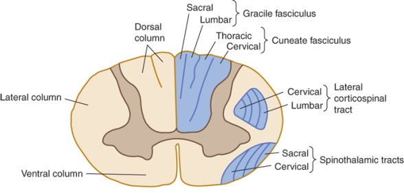
These neurons project from the body to the spinal cord. Then they travel up one of two ways. The medial lemniscus tract is split into the gracile and cuneate fasciculus. If the nerve is located at the lower limb, it travels through the gracile fasciculus. If the nerve is located in the upper limb, then it travels via the cuneate fasciculus. To see where these two tracts are located within the medial lemniscus tract, the image below has them labeled. Remember, the spinal cord is symmetrical, so the same division applies to the other side as well.
Now we can follow the tracts to the brain (even though technically we’re only talking spinal cord). Like the corticospinal tract, this tract passes through the medulla. In this case however, there are two nuclei (a collection of cell bodies, somewhat, but different from the psudounipolar neuron we just discussed) where the dorsal column synapses. These two nuclei are named for the tract they are associated with, they are the cuneate nucleus and gracile nucleus. Since these are the second neurons that are involved in this pathway, we call them second order neurons.
Once they synapse on their respective nucleus, they decussate at the internal arcuate fibers. This is named for the way they look, in a cross section they “arc” across the brainstem. Scientists aren’t super creative when it comes to naming, but trust me this is a good thing. Let’s look at that cross section so you can see what I’m talking about.
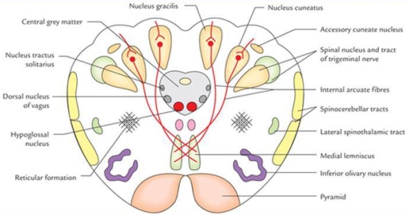
The internal arcuate fibers (red) and the nucleus gracilis and cuneatus labeled accordingly.
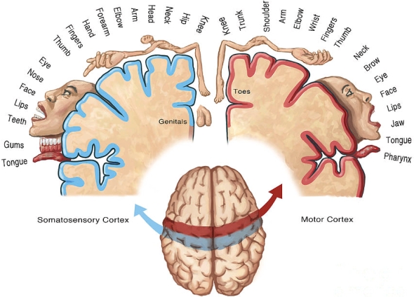
Once they travel up the brainstem, we refer to them as the medial lemniscus. These then synapse on a nucleus at the thalamus called the ventral posterolateral nucleus (this tells us the location, ventral, posterior, and lateral and again we are not creative with names and that is good). This nucleus sends projections (now third order since these are the third neurons to be involved in this chain) to the postcentral gyrus in the cortex. You may remember we called this portion of the cortex the somatosensory cortex. If you need a refresher about the location of the somatosensory cortex, below you can see the somatosensory cortex (blue) sits directly behind the motor cortex (red).
So now we’ve covered the entire pathway! There are only three neurons that make up this pathway. The first is in the dorsal root ganglion, the second in the cuneate nucleus, and the third in the ventral posterolateral nucleus. If you’re wondering, that makes some of these neurons extremely long (at least in the way I tend to think of neurons).
Next up, I’m actually not sure which tract I want to cover tomorrow. Maybe the posterolateral tract, also known as the tract of Lissauer, named for the guy who found it. It’s my FAVORITE tract (because I’m a nerd like that) and it is particularly weird about how it’s organized (in my opinion anyway).
Until next time, don’t stop learning!


I love this!!
LikeLike
June 6, 2020 at 3:45 am
Thank you!
LikeLike
June 6, 2020 at 9:39 am