Know your spinal cord – The anterior white commissure
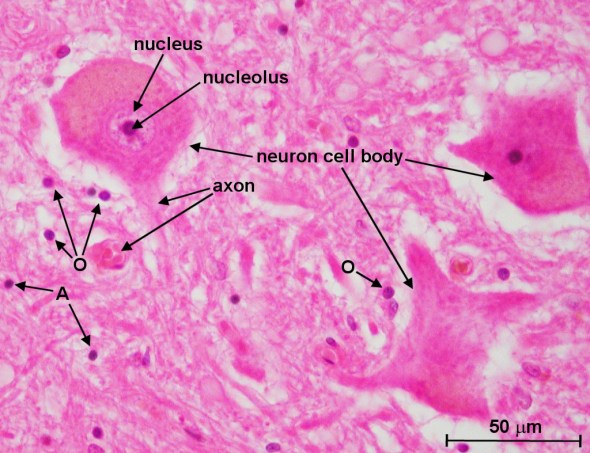
Cross section through human spinal cord (lumbar region, L1) showing motor neurons. Astrocytes and oligodendrocytes are labeled ‘A’ and ‘O’, respectively. The objective magnification used is 40x.
Welcome to day thirty of knowing your spinal cord. I feel like that is a lot of spinal cord knowledge for just covering the basics. In any case, if you’re just finding us, welcome! I’ve created a whole new neuroanatomy category just for these posts so you can find them easy and they are in reverse chronological order. Is the anterior white commissure a tract of the spinal cord? Well not really, but it does have an important job and we keep referencing it, so let’s talk about what it does exactly.
If we recall, the spinal cord is roughly the diameter of your pinky. It’s essentially a noodle brain™ and for that reason, every structure has a critical role. That is why we are covering the anterior white commissure today. It’s role is in spinal signaling, but falls somewhere between the neural circuitry of the grey matter and the tracts of the spinal cord. However, just like the grey matter and white matter tracts, the anterior white commissure has a defined structure that we can discuss.
When we talk about the anterior white commissure, we are talking about a collection of nerve fibers, making it a pseudo-tract of the spinal cord. Instead of relaying information to the brain from various sensory modalities, the anterior white commissure decussates (crosses the midline) in the spinal cord to relay information from or to the contralateral side of the brain.
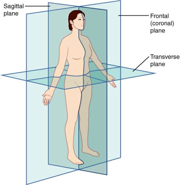
The coronal plane is a frontal plane to the body. The sagittal plane defines the side. The transverse plane is a plane that cuts through the body dividing the upper and lower halves of the body
Located directly behind the anterior median fissure, it is present the full length of the spinal cord. While white matter tracts run parallel to the sagittal and coronal planes of the body, the anterior white commissure runs parallel to the transverse plane. Above you can see an image defining the planes of the body. While we don’t consider it a tract of the spinal cord, there are similarities in its function and it can analogously be thought of as a tract relaying information to the contralateral side of the spinal cord. Below is an image showing the anterior white commissure. The large white spot is the anterior median fissure, a split in the cord, directly above that (where the arrow is pointing) is the anterior white commissure, in the image below you can actually see the nerve fibers that decussate in this region.
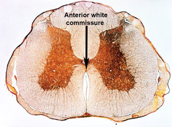
The anterior white commissure (labeled) is located directly between the anterior median fissure and the grey commissure (the dark region that connects the left and right side forming the butterfly shape)
Among the important pathways that decussate in the spinal cord via the anterior white commissure are the second-order neurons of the spinothalamic tract and the upper motor neurons of the anterior corticospinal tract. This means that two out of the five sensory modalities, pain and temperature, cross sides at the anterior white commissure, reaching the contralateral side about two vertebral levels rostral (above) to their origin. These crossing fibers make the anterior white commissure an important link in communication between the brain and the contralateral side of the body for both sensory and motor pathways.
Now that we’ve covered the anterior white commissure, there are still some anatomical features that we should probably cover. Then we can discuss some of the research that I am doing and my motivations for studying the spinal cord. It will be an interesting week or so for sure!
Until next time, don’t stop learning!


But enough about us, what about you?