Know your spinal cord – Astrocytes

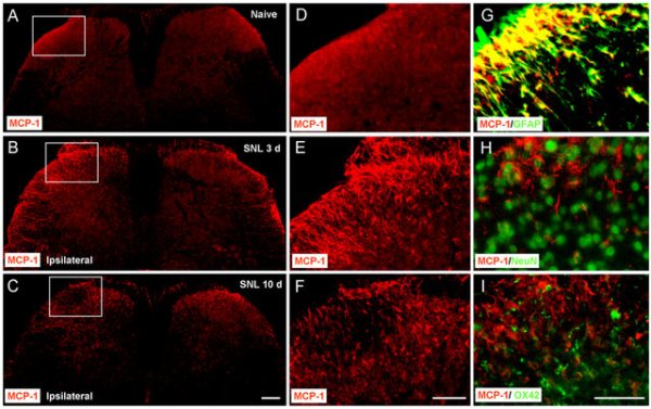
SNL induces MCP-1 upregulation in spinal cord astrocytes (A–C)MCP-1 expression in the spinal cord of naïve animals (A) and SNL animals at 3 days (B) and 10 days (C). Scale bar, 200 μm. (D–F) High magnification images of (A–C), indicated in the white boxes of A–C, show the dorsal horn of the ipsilateral spinal cord. Scale bar, 100 μm. (G–I) Double staining shows that MCP-1 is colocalized with GFAP, a marker for astrocytes (G), but not with NeuN, a marker for neurons (H) or OX42, marker for microglia (I). Scale bar, 50 μm. DOI: 10.1523/JNEUROSCI.3623-08.2009
Day forty-nine in the spinal cord series! You can find all the posts in this series in our super useful neuroanatomy category. A couple of posts back we introduced glial scarring, one of the problems we need to overcome to help people with spinal cord injuries. That led to the realization that we needed to introduce the glial cells, so yesterday we covered the oligodendrocytes and today we are talking about the astrocyte. Now that we have some background of how we got here, let’s introduce today’s topic.


