Know your spinal cord – Astrocytes
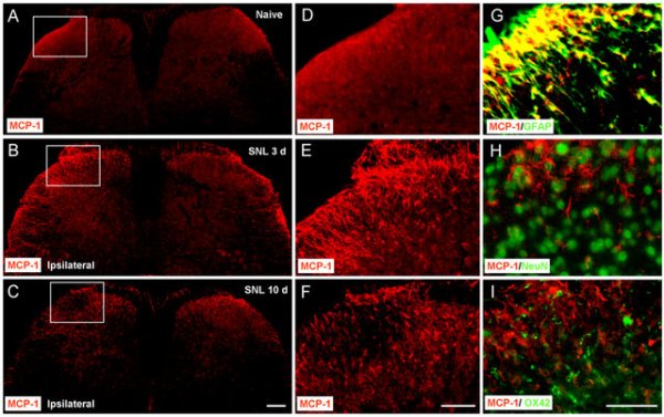
SNL induces MCP-1 upregulation in spinal cord astrocytes (A–C)MCP-1 expression in the spinal cord of naïve animals (A) and SNL animals at 3 days (B) and 10 days (C). Scale bar, 200 μm. (D–F) High magnification images of (A–C), indicated in the white boxes of A–C, show the dorsal horn of the ipsilateral spinal cord. Scale bar, 100 μm. (G–I) Double staining shows that MCP-1 is colocalized with GFAP, a marker for astrocytes (G), but not with NeuN, a marker for neurons (H) or OX42, marker for microglia (I). Scale bar, 50 μm. DOI: 10.1523/JNEUROSCI.3623-08.2009
Day forty-nine in the spinal cord series! You can find all the posts in this series in our super useful neuroanatomy category. A couple of posts back we introduced glial scarring, one of the problems we need to overcome to help people with spinal cord injuries. That led to the realization that we needed to introduce the glial cells, so yesterday we covered the oligodendrocytes and today we are talking about the astrocyte. Now that we have some background of how we got here, let’s introduce today’s topic.
Astrocytes are star shaped glial cells. The comes from the latin and basically translates to star cell. Frankly these are so glial cells are so complex it’s hard to give a good overview, so like yesterday I think today we will focus on some of the main functions. Remember, this is an introduction, you could spend years learning about astrocytes alone and still not know everything. Below is a rendering of what astrocytes look like, we will look at actual images of them in a moment, but this gives a good idea of the shape.
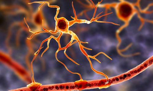
Astrocyte attached to a blood vessel
Astrocytes could very well be the most numerous cells of the vertebrate central nervous system. It’s hard to count them all you see, but we do a pretty good job of estimating, so it’s a pretty good guess that there are quite a few. The work they do is just as diverse as their numbers. Primarily thought of as giving the brain it’s structure, astrocytes are also found in the spinal cord. They aren’t passive though, they also play an active role which includes the secretion or absorption of neural transmitters and maintenance of the blood–brain barrier. In the dorsal horn of the spinal cord, they respond to almost all neurotransmitter and, upon activation, release a multitude of neuroactive molecules such as glutamate, ATP, nitric oxide (NO), and prostaglandins (PG), which in turn influences neuronal excitability. Below is an actual transverse slice of spinal cord tissue showing the astrocyte concentration (stained green).
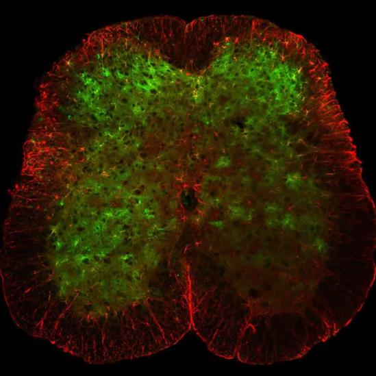
Astrocytes (green) in a transverse slice of spinal cord. DOI:10.1038/ncomms11450
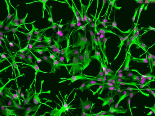
They also have a role in the provision of nutrients to the nervous tissue, maintenance of extracellular ion balance and a role in the repair and scarring process of the brain and spinal cord following traumatic injuries. The proportion of astrocytes in the brain is not well defined; however, depending on the counting technique used, studies have found that the astrocyte proportion varies by region and ranges from 20% to 40% of all glia! Below is another image showing human astrocytes under a microscope. They really do look like the rendering we showed above.
Now, upon injury to the spinal cord, astrocytes fill up the space to form a glial scar, and may contribute to neural repair. The role of astrocytes in CNS regeneration following injury is not well understood though. As we mentioned in the glial scarring post, the glial scar has traditionally been described as an impermeable barrier to regeneration, thus implicating a negative role in axon regeneration. However, recently, it was found through genetic ablation studies that astrocytes are actually required for regeneration to occur. More importantly, the authors found that the astrocyte scar is actually essential for stimulated axons (that axons that have been coaxed to grow via neurotrophic supplementation) to extend through the injured spinal cord.
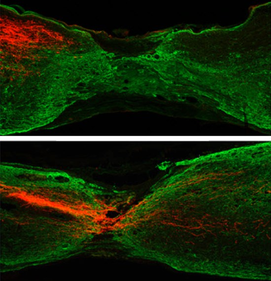
HKUST researchers cut mouse corticospinal tract axons (labeled red). A year later, they deleted the Pten gene in the experimental group (bottom) but not the control group. The Pten gene removal resulted in axon regrowth in seven months, unlike the control group (top). DOI: 10.1523/JNEUROSCI.3637-14.2015
Basically, we know they perform an important role in the CNS and for us, within the spinal cord itself. However, we don’t quite understand if the astrocytes are helping or hindering spinal cord regeneration. More work needs to be done, but in mouse models researchers have shown that it is possible for regeneration to occur and that astrocytes play a key role in this. The image above (top) shows glial scarring of a mouse spinal cord (green), along with a treated mouse where the axon (red) has grown through the scar tissue (bottom). Does the same thing apply to humans? We’re not sure yet, but it is exciting to see the possibility of regenerating the spinal cord tissues after injury.
That is your crash course into astrocytes. It’s just a small look at all the things they do, but one that (hopefully) helps us make more sense of what glial scarring is and why it may not be a completely bad thing. We have two more types of astrocytes to talk about, so tomorrow we will be introducing the ependymocyte. Not quite as complex as the astrocyte (that we are aware of anyway), it plays an important role in the spinal cord and tomorrow we will see what that role is!
Until next time, don’t stop learning!


But enough about us, what about you?