Know your spinal cord – Ependymocytes
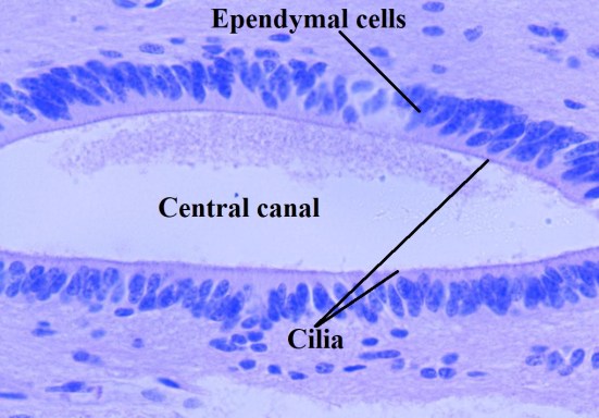
Ependymal cells, which create cerebral spinal fluid (CSF), line the ventricles of the brain and central canal of the spinal cord. These cells are cuboidal to columnar and have cilia and microvilli on their surfaces to circulate and absorb CSF.
We made it to yet another milestone, day fifty in our know your spinal cord series! As usual, you can find each and every one of these posts neatly organized in reverse chronological order using our neuroanatomy category. For the past couple of posts, we’ve introduced the types of glial cells, probably a bit poorly, but they are just so complex we can only really focus on a few of the functions. Needless to say they are very important cells. Today we are talking about the third (of four) types of glial cells found in the spinal cord (and brain), that is the ependymocyte. Let’s take a look.
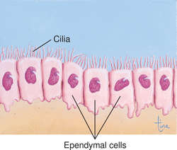
The first and (probably) main function of ependymocytes is the formation of ependyma (aptly named). These cells line the CSF-filled (cerebrospinal fluid) ventricles in the brain and the central canal of the spinal cord. Embryonic ependymal cells are ciliated (have cilia) and some retain their cilia permanently. Cilia are tiny hair like projections that move in a rhythmic motion to push fluid in a particular direction. For example your nose, trachea, and lungs are lined with them and help move mucus around. Below is an image showing what cilia of the ependymal cells look like.
Ependymal cells lining the central canal of the spinal cord have cilia, hair like projections that move CSF through the canal.
In the case of the ependymal cells in the spinal cord, they push cerebrospinal fluid (CSF) through the canal and help keep it moving. They have a very distinct pattern to them that helps push the fluid. There are types of disease that affect this movement, but we won’t cover that. Instead let’s look at what the healthy movement looks like. Below is a short animation showing the movement pattern of the cilia in healthy cells. You can see the cells work in a coordinated fashion to push the fluid in a particular direction.
Now that we’ve explained cilia, let’s get back to the ependymal cells. Moving fluid and forming the ependyma isn’t the only functions they perform. They also are responsible for maintenance of homeostasis in the brain. This shouldn’t be much of a surprise since they form a barrier, because we haven’t shown it yet, below is an image of the barrier the ependymal cells form in the central canal, this is a schematic view for an adult mouse, but the human version looks remarkably similar.

Schematic drawing of the adult mouse ependymal region. Adapted with permission from Sabourin et al (32) and Hamilton et al (21). DOI: 10.2741/3734
Here’s where things get interesting and take an odd tern. Up until now, the functions are pretty much expected given the structure of the cell, right? Well, they also have a role in spinal cord regeneration, or I should say they might. In adulthood, the spinal cord stem cell potential is restricted to ependymal cells. Ependymal cells are activated by traumatic SCI, self-renew and differentiate into astrocytes and oligodendrocytes. Moreover, when the proliferation of ependymal cells is impaired, the formation of the glial scar after SCI is heavily compromised, detrimentally affecting neuronal survival.
So once again we are left with a very complex role in the spinal cord for a type of glial cell. The odd thing about this line of research is the confusion surrounding the ependymal cells (glial cells in general really). They don’t quite qualify as stem cells because once they differentiate the reserve numbers, that is the number of cells that remain undifferentiated, don’t replenish. Further exacerbating things, as we get older the reserve number appears (key word here is appears since this is all still very new research) to drop and the level of injury severity to which they respond increases.
Most of this research is limited to mouse/rat models however, so it’s hard to say how this will translate to humans. The important thing to remember here is that these cells are yet another important factor in spinal cord regeneration and repair. Once again I have to leave you with more questions than answers because this research is still ongoing and we honestly don’t know a lot about what functions they have and how that changes as we age.
Next up is our fourth and final glial cell of the spinal cord (and brain, but we only really are interested in the spine… right?) the microglia. I wish I could say that they have a simple function that I could concisely explain, but guess what? They do not. If anything these few posts should highlight the fact that glial cells are absurdly complex and have a lot of functions (some of which we may not even know). But let’s save that for the next post.
Until next time, don’t stop learning!


But enough about us, what about you?