Know your spinal cord – The spinal nerves
Here we are, a week into knowing your spinal cord (remember we have a new category for you to find these posts). If you’re just starting out, you may want to look at our new neuroanatomy category and start with the first post. For those of you who have been following along, we covered some of the major tracts of the spinal cord, so let’s dive into the structure some! First up, let’s talk about spinal nerves and what exactly these guys do.
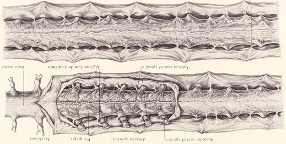
We’ve talked about them in the past few posts, spinal nerves. Specifically we discussed that there is something called the dorsal root ganglion, which inside of that houses the cell body of the nerve. This nerve is a pseudounipolar neuron, which is named after the way it looks. Below is an image of the dorsal root ganglion and the pseudounipolar neuron.

Notice that in our dorsal root ganglion there is a cell body, nerve guy is our pseudounipolar neuron and the position of the cell body is why it is a pseudounipolar neuron (because it is away from the axion).
You’ll also notice we have a ventral root as well. The dorsal root is the afferent pathway and the ventral root houses our efferent pathway (remember Efferent signals Exit the brain). You may have also caught that unlike the dorsal root, there is no ganglion and there is no pseudounipolar neuron. That is because the cell body sits inside the spinal cord (see the red and blue blob). This type of neuron is called a multipolar neuron. If you’re interested, there are a couple other types (including a unipolar, not just the pseudounipolar) and we have a nice image below that shows them all.

Like the vertebra of the spinal cord, we number the nerves that come off the spinal cord in accordance to where they exit (so cervical, thoracic, lumbar, sacral, and coccygeal. Our spinal cord is (for the most part) a mirror image of itself, so when we talk about nerves, we are really talking about pairs of nerves (not to be confused with the dorsal and ventral roots, we are talking about the left and right side of the spinal cord not front to back). That being said, we have 31 pairs of spinal nerves, they are:
- 8 cervical nerves (located in the neck)
- 12 thoracic nerves (located in the chest)
- 5 lumbar nerves (located in the abdomen)
- 5 sacral nerves (located in the pelvis)
- 1
lonelycoccygeal nerve (located in the tailbone)
Keep in mind it isn’t just one nerve that exits or enters, it is a cluster of nerves. The number of axons (or nerve fibers) in a the dorsal and ventral roots can vary anywhere from just a handful, to more than a million!
Now that we’ve covered that, we can also talk about how they innervate the body. While the spinal cord (much like the brain) is a black box, we have a really good handle on how it connects to the body. If you’ve never seen one, every single drawing looks like someone who’s never seen a human and had to find a way to pose them. Below is a good example of this, while it’s funny looking, it actually does a great job of showcasing where the nerves attach to the body. From a diagram like this, you can actually determine approximately where you would lose sensation if there were a spinal cord injury.
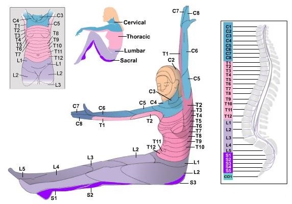
These maps are commonly called dermatome maps. A dermatome is a fancy term for the an area of skin supplied by peripheral nerve fibers originating from a single dorsal root ganglion. This means that if we were to cut a nerve, you would lose sensation from that dermatome. This is a sensory map, so it highlights the ascending pathway. Derma, meaning skin is the giveaway for this map.
Knowing that, it may not surprise you that we can do the same for the descending pathway (motor pathway) and it is called a myotome map (myo meaning muscle). Interestingly the myotome map looks very similar (but not exact, a good example of this is the hand being innervated at C6 below and C7 above) to the dermatome map. So let’s look at the myotome map. This one is posed a little nicer.

So now we have a very detailed map of where everything connects! Not only that, but we have a map of both the major afferent pathway and efferent pathway. So once again, while we don’t know exactly what’s going on in the spinal cord, we do have a very good idea of how it all attaches at least.
Where do we go from here? Good question, I think, THINK, that I will cover some of the grey matter organization next. We’ve done a good job of looking at the white matter organization, but we haven’t really touched on the grey matter. This will give us a more rounded knowledge base I think. We will also (probably) go over reflexes, what we know about the spine, what we’re trying to figure out, and some of the more interesting cases of spinal cord injury.
So that’s your look ahead for the day. I hope by the end of this post, you’ve gained an understanding of how the nerves that connect to the spinal cord are organized and what they do. If you know that (even in the broadest sense) then I’ve done my job. If there are any questions, comments, or concerns, don’t hesitate to ask!
Until next time, don’t stop learning!


But enough about us, what about you?