Know your spinal cord – The grey matter
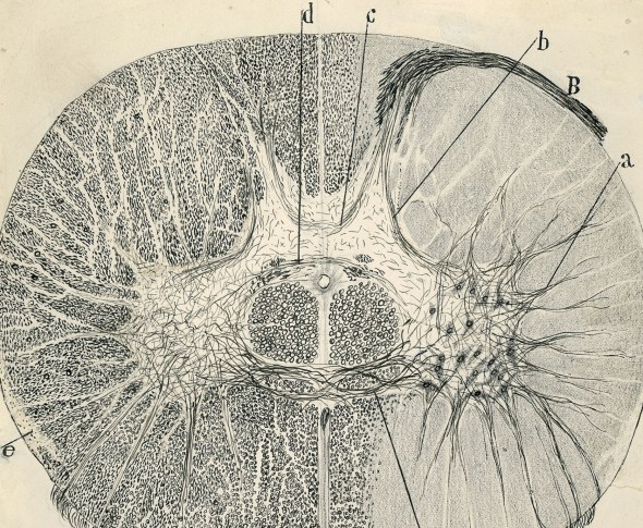
Drawing by Ramon y Cajal, a famous Spanish neuroscientist.
Today is day eight! I can hardly believe it, but here we are, day eight of spinal anatomy. For those of you who are just joining us, we have a whole new category just for these posts and they are in order from newest to oldest, so start at the bottom and work your way up. For those of you who have been following along, today we are tackling the grey matter of the spinal cord, a somewhat complex region where all the action takes place.
From our current view of neuroanatomy, the grey matter is where all the processing happens. While white matter sends data (like a cable) the grey matter is more like your CPU, taking all that information and transforming it somehow. The brain has the grey matter on the outside and the white matter on the inside, however the spinal cord has the reverse. This is likely due to the surface area increasing with the radius and the spinal cord for all its intelligence primarily relays information to and from the brain.
Some of this we’ve touched on in previous posts, because it’s hard not to include other systems when we’re talking neuroanatomy. However, let’s take it from the broadest sense. If we look at a cross section of spinal cord, we see a white matter exterior, with a grey matter interior, which is in the shape of a butterfly. We call each of the protrusions a horn of the spinal cord and depending on where you are in the cord, we have three of them. The dorsal horn, lateral horn, and ventral horn, which can be seen in the image below.
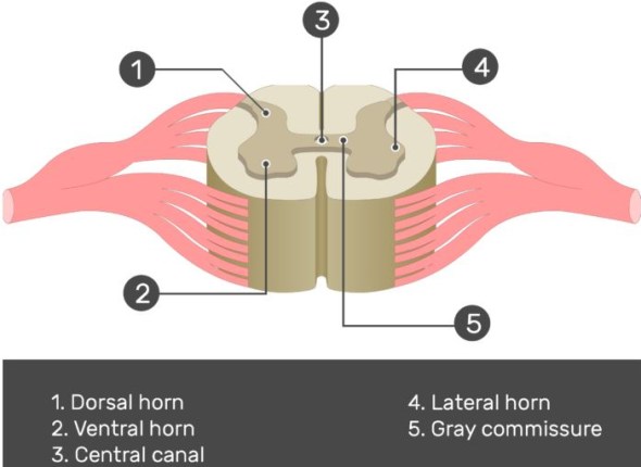
There are three “horns” of the spinal cord. The dorsal horn (1), the lateral horn (4), and the ventral horn (2).
While the dorsal horn and ventral horns run the full length of the spinal cord, the lateral horn does not, it is located in the thoracic and upper lumbar regions of the cord exclusively. This is because the lateral horn innervates visceral and pelvic organs so it is not needed the full length of the cord like the dorsal and ventral horns. More specifically it is part of the sympathetic nervous system and receives input from brain stem, organs, and hypothalamus. This makes it an important part of the spinal cord (even if it doesn’t play as prominent a role as the dorsal and ventral horns).
Here’s where things get tricky! We have two different ways of looking at the grey matter, we can organize it by nuclei, which was how it was originally organized. However, in the 1950’s a guy named Bror Rexed offered a different organization called the Rexed Laminae (or layers). He identified these layers, or again laminae, within the spinal cord where cells were grouped according to their structure and function, rather than solely on location. So we have two ways to represent this. To make things more complex, if we organize by nuclei, we have six different structures, if we go by laminae, we have ten. For completeness, we will cover both!
Let’s start with the nuclei. As I mentioned, there are six main structures in this way of organizing the spinal cord, they are:
- Marginal zone (MZ, posterior marginalis) – this is located at the tip of the dorsal horn, it handles relaying pain and temperature sensation to the brain.
- Substantia gelatinosa (SG) – we talked about this previously, if you recall it is located at the top of the dorsal horn, the SG is important for relaying pain, temperature and light touch sensation to the brain.
- Interomediolateral nucleus (IMN) – located in the intermediate column and lateral horn, the IMN relays sensory information from viscera (internal organs) to the brain, and autonomic signals from the brain to the visceral organs.
- Dorsal nucleus of Clarke (DNC) – the most dorso-medial nuclei, the DNC relays unconscious proprioceptive information to the brain. This area is only found in spinal segments C8 to L3.
- Lateral motor neurons and medial motor neurons (MNs) – you may recall we covered this briefly as well, it is located in the ventral horn. Composed of motor neurons that innervate visceral and skeletal muscles.
- Nucleus proprius (NP) – located adjacent to the substantia gelatinosa in the dorsal horn, the NP relays mechanical and temperature sensation to the brain.
Don’t worry, we will look at an image of where these are located in just a moment. Typically images show both nuclei and laminae organization so for now, these are the six, their function, and location.
Now let’s talk about the laminae of the spinal cord. They are numbered 1-10 and have a very clear organization, so you will know where 1 is vs 10 because they actually go in an order. Like the nuclei, we can list the location and the function, becuase there are a couple we will change the way we make this list and use subheadings and bullets this time.
Lamina I
- very tip of the dorsal horn (similar to the marginal zone)
- these cells respond to noxious or thermal stimuli
- sends information to the brain by the contralateral (opposite side) spinothalamic tract
Lamina II
- Involved in sensation of noxious and non-noxious stimuli, and modulating sensory input to contribute to the brain’s interpretation of incoming signals as painful, or not.
- Sends information to Lamina III and IV (which we will cover below)
- Located similarly to the substantia gelatinosa
Lamina III
- Involved in proprioception and sensation of light touch.
- Cells in this layer connects with cells in layers IV, V and VI.
- This is an oddball lamina, but it partially corresponds to nucleus proprius
Lamina IV
- Involved in non-noxious sensory information relay and processing.
- Cells connect with those in lamina II (see above)
- Again, an oddball which partially corresponds to nucleus proprius
Lamina V
- Relays sensory, including nociceptive (potentially painful), information to the brain via the contralateral (opposite side) and spinothalamic tracts
- Receives descending information from the brain via the corticospinal and rubrospinal tracts.
Lamina VI
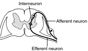
- Contains many small interneurons (image below showing what an interneuron does) involved in spinal reflexes
- Receives sensory information from muscle spindles (involved in proprioception).
- Sends information to the brain via ipsilateral (same side) spinocerebellar tract.
Lamina VII
- Large, heterogenous zone that varies through the length of the spinal cord.
- Receives information from Lamina II to VI, and from viscera (internal organs)
- Relays motor information to the viscera
- Cells involved in the autonomic system
- Dorsal nucleus of Clarke is part of Lamina VII
Lamina VIII
- Varies depending on spinal cord level, but is most prominent in cervical and lumbar enlargements
- Cells are involved in modulating motor output to skeletal muscle
Lamina IX
- This guy depends on the level you are looking at because the size and shape varies between spinal cord levels
- Distinct groups of motor neurons that innervate skeletal muscle.
Lamina X
- Surrounds the central canal – the grey commissure
- Axons decussate (cross over) from one side of the spinal cord to the other
So as you can see, some of the laminae correspond to the nuclei organizational method, but not quite. Let’s clear this up a bit by showing both the nuclei and laminae organization in one figure.
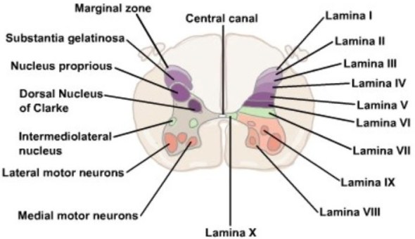
That is a lot of information for sure, but I hope that it all made sense. A helpful way to remember the function of the lamina is this, laminae 1-4 (I-IV) are predominantly involved in interpreting and relaying sensory information from the body to the brain. Laminae 5-6 (V-VI) handle proprioceptive sensation and act as a relay between the periphery to the midbrain and the cerebellum. Laminae 7-9 (VII-IX) form the final motor pathway to initiate and modulate motor activity. Lastly Laminae 10 (X) is our decussation laminae primarily moving data between the left and right side of the cord.
We can make it even simpler and say neurons in the laminae towards the dorsal portion of the cord (IE: laminae one through five) are handle sensory information from the body to the brain. Neurons in the laminae towards the ventral portion of cord (IE: laminae seven, eight, and nine) are involved primarily in executing movement and controlling the functions of the bodies organs.
“Put even more generally, the dorsal portion of the grey matter deals with sensory information while the ventral portion deals with movement.”
You may notice lamina ten (X) is sort of the odd lamina out. Someone had to have the job of decussation and it was this lamina that got it. Keep in mind that this is just our understanding of the lamina as well, they could have other jobs we don’t know, do things we don’t know, or innervate areas that we haven’t seen yet. Our understanding of the spinal cord (and indeed the human body itself) is always changing as we discover new and interesting things about it. However, as of this writing, those are the jobs of the lamina and nuclei as we understand them.
This was quite the post, next I think we need to cover some of the minor tracts, not being able to link out to them here was somewhat annoying. Those posts will be short becuase the tracts are very specific, but it would be nice for me since each of these posts takes upwards of 2-3 hours despite having the knowledge to write it.
Until next time, don’t stop learning!


But enough about us, what about you?