Know your spinal cord – The spinal cord enlargements
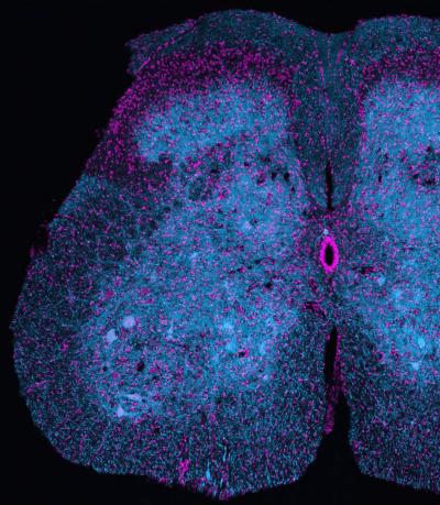
Mitochondria in a section of spinal cord
Here we are at day thirteen of knowing your spinal cord! As always, we have a whole special category for these posts called neuroanatomy and if you’re not after a specific topic, I recommend starting at the medullary pyramids. Today’s post is about something important that we haven’t touched on very much, the cervical and lumbar enlargements of the spinal cord, so let’s get started.
We have not one, but two enlargements to the spinal cord. These enlargements hold the circuitry that control the upper limbs and lower limbs. This is the reason they are enlarged areas of the spinal cord, becuase there is a higher amount of grey matter in these areas than other locations along the cord. Let’s look once again at how the spinal cord changes as we travel across the spinal cord.

We’ve used this image before, but as you can see from above, depending on where we are in the spinal cord, we have a different amount of white to grey matter. As we’ve already seen grey matter is where processing happens and is analogous to your computer’s CPU. On the other hand, white matter sends and receives data, sort of like an HDMI cable. Now let’s talk about the cervical enlargement and then we can get into the lumbar enlargement.
The cervical enlargement is responsible for coordination of the upper limbs. It starts about C5 and ends at approximately T1. Because we denote spinal levels based on the vertebrae number, this is an approximation, but it is fairly close. The cervical enlargement is roughly 38mm in circumference making it one of the largest portions of the spinal cord (the largest in the spinal cord proper). Because we need to be dexterous and as we’ve already covered in our CPG post and our review of a paper on spinal stretch reflexes the neural circuitry that controls the upper limbs is very complex.
The lumbar enlargement on the other hand is responsible for coordinating the lower limbs. This enlargement starts at roughly T11 and ends at approximately S2 (we haven’t covered what S stands for, but it is sacral from the sacrum and you can see where this is located in the above image). As with the cervical enlargement we have (probably) CPG and spinal stretch reflex neural circuitry housed here. This enlargement is slightly smaller than the cervical enlargement at roughly 33 mm in circumference.
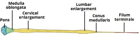
Below is another image showing the anatomy of the spine in relation to the cervical and lumbar enlargements. There are a couple of things we haven’t talked about in detail. The conus medullaris is the term for the end of the spinal cord and the filum terminale is a strand of fibrous tissue that helps anchor the spinal cord. We covered both of these briefly in the spinal anatomy post and we may go into more detail, but for now just know they exist.
The nice thing about the enlargements is that the neural circuitry is nice and contained. If it were more distributed spinal cord stimulation would (probably) not be as therapeutic. If you haven’t and want to see a video showing how spinal cord stimulation helps restore function, at the end of our central pattern generator post yesterday we have a nice little video that shows the before and during effects of stimulation.
Another nice feature of the cervical and lumbar enlargements is that we can specifically stimulate one or the other (even both). Additionally, we know that they “talk” to each other. At our roots we were quadrupeds and even now we coordinate our upper limbs with our lower limbs when we walk (arm swing). One current area of research is trying to determine how exactly they connect and the circuitry that drives one or the other. There has been some invasive animal studies along with invasive human, and non-invasive human studies that are trying to work this out even now.
What we are interested in is determining how an input to the cervical enlargement changes a response to the lumbar enlargement and vice versa. Do they use the same afferent and efferent pathways? Does cervical enlargement activation change lumbar activation? Does lumbar activation change cervical activation? These are just some of the questions we are trying to answer and finding this out will help us understand spinal cord injury and how we can better treat it.
Next up I think we will cover the cauda equina. Why it exists is an interesting story and while it doesn’t have a specific function like the lumbar or cervical enlargements it is an interesting part of the anatomy of the spinal cord.
Until next time, don’t stop learning!


But enough about us, what about you?