Know your spinal cord – The F-wave
It’s day thirty-nine of our know your spinal cord series and we’re only touching the surface (so to speak)! If you’re just joining us, then welcome! You can find all of our spinal cord knowledge in the handy neuroanatomy category. Well as these things typically happen, yesterday brought up an interesting gap in our knowledge base. While I introduced the H-reflex, we never talked about the F-wave! So of course yesterday’s post probably left some of you scratching your head as to what an F-wave even is, fear not we’re going to clear that up today!
If we are (eventually) going to talk about my research we need to talk about the tools we use to probe the spine. Electrical stimulation is super nice because we can precisely control the duration, pulse width, amplitude, and the phase characteristics (like a monophasic or biphasic pulse), just to name a few things! This means consistency and if we are going to try to find the secrets of the spinal cord, we need to be able to be consistent with our testing.
The Hoffman reflex (H-reflex) named after the guy who found it (of course), is one way we probe the spinal circuitry. In that case we stimulate our 1a afferent and are rewarded with a reflex response shortly after. We also introduced the M-wave in that post, or muscle response. If we cranked up the amplitude on the pulse we sent, eventually we would stop stimulating the spinal circuitry and be left with just a response from the muscle.
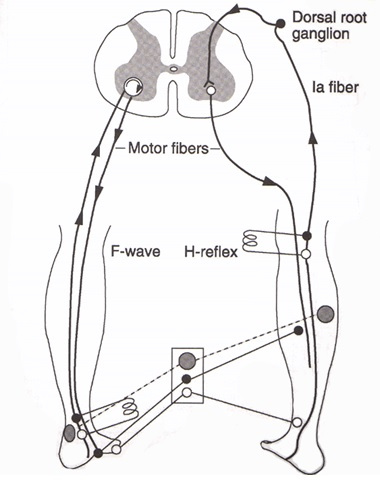
So how is the F-wave different from the H-reflex or the M-wave? Well it just so happens I have a super fancy graphic to explain it, but first let’s talk a little bit about what the F-wave is all about. Unlike the H-reflex, the F-wave was named after the foot (seriously), because that is where researchers were stimulating when it was found originally. The F-wave is hard to record and is best done with the smaller distal muscles (such as the hand or like its namesake, the foot).

To understand the F-wave we need to talk about how nerves transmit data, above is the anatomy of your typical neuron. When a neuron fires it creates something called an action potential. An action potential is where the membrane potential of a specific cell location rapidly rises and falls, this change in membrane potential propagates all the way down the axion. It’s easier to see this in action and below is an animation showing how an action potential occurs. Now one important thing to notice, the action potential is one way. It starts at the cell body (soma) and ends at the axon terminal, this is always the case with this type of cell (remember we have other types, but since this is high level, just know we are simplifying things a bit).

This brings us to the F-wave. To create an F-wave we apply a strong (supramaximal) electrical stimuli to the distal (distal means away so your hand is distal to your elbow) portion of a nerve. This causes an antidromic impulse, antidromic is the fancy term we use when it travels counter to the normal action potential, which we refer to as orthodromic. Basically we now have a pulse traveling from the axon terminal to the cell body in the spinal cord along the alpha motor neuron. This causes the neuron to backfire and create an action potential in the normal direction. This doesn’t happen in nature, but we can sort of force it. This is also why the F-wave is tricky and works best with smaller muscles, because of the amplitude you need to generate the response. As you might imagine this also generates a rather large M-wave. However, we can easily separate the M-wave from the F-wave from the latency.
Now that we’ve introduced how it is created and what is happening, let’s look at the graphical representation of the M-wave, H-reflex, and F-wave. This will help make sense of everything, but it also explains why we use the F-wave to probe the spinal cord.

Researchers use the F-wave because it gives a very targeted response from a very small motor pool. This is useful because in spinal cord injury, these motor pools may not even be connected, but we can still use the nerve to stimulate them and measure the response. While this technique is more used for nerve conduction studies, studies like the one I reviewed yesterday highlight the versatility of the technique in understanding spinal cord injury.
That is it for your crash course on the F-wave! Hopefully this has cleared a few things up and helps make yesterdays paper more accessible to you. Now that you’ve gotten an introduction to the H-reflex, M-wave, and F-wave, we can introduce another type of stimulation… but you’ll have to wait until tomorrow to read about that.
Until next time, don’t stop learning!


This is a great ppost
LikeLike
February 14, 2023 at 12:21 am
Thank you!
LikeLike
February 19, 2023 at 11:28 am