Day 196: Review – Changes in Motoneuron Excitability during Voluntary Muscle Activity in Humans with Spinal Cord Injury
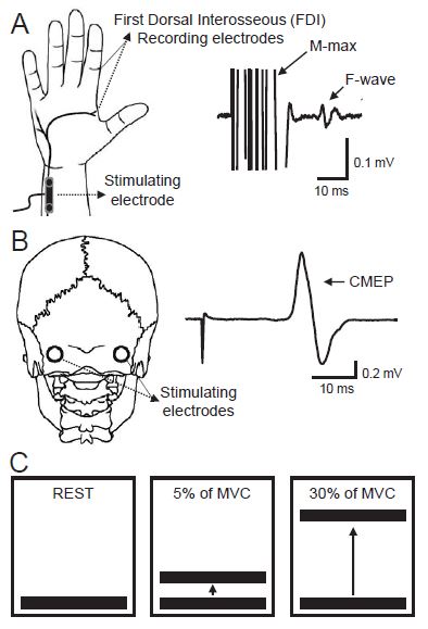
Figure 1. Experimental setup. A, Schematic representation of the hand showing the ulnar nerve and F-waves recorded from the first dorsal interosseous (FDI) muscle. B, On the left side, schematic representation of the head showing the electrodes placed at the cervicomedullary junction and on the right side a raw trace showing a cervicomedullary motor evoked potential (CMEP). C, A cartoon showing the concept of the visual feedback. Individuals were tested at rest (left single horizontal line) and during 5% (middle double horizontal lines) and 30% (right double horizontal lines) of maximal voluntary contraction (MVC).
A little detour from our spinal cord series for my fourth critical review paper. As usual, my PI get a copy and so do all of you! You can read my first looking at elbow spinal stretch reflexes here. My second where I review modulating spinal cord excitability with a static magnetic field here. Or the third where I review modulating the H-reflex while walking in spinal cord injury populations. Today is an interesting paper on motoneuron excitability while walking in spinal cord injury populations. It’s a really cool paper, so here’s my review.
Current evidence shows that after a spinal cord injury (SCI), resting motoneuron excitability increases. It has also been shown that humans with SCI have exaggerated tendon reflexes along with decreased inhibition in spinal neuronal signals, and alteration in group 1a projections to motoneurons with respect to healthy individuals. However, in humans with SCI, motoneuron excitability change during voluntary muscle activity is poorly understood.
This study examines the amplitude and persistence of F-waves and size of cervicomedullary motor evoked potentials (CMEPs). Researchers recruited sixteen individuals with chronic incomplete SCI (≥ 1 year) who had cervical injury (C2-C6) and eighteen age-matched controls. Electromyographic (EMG) recordings were taken from the first dorsal interosseous muscle (FDI) on the right side in the controls and the less affected hand in the SCI subjects. Participants were sat in a chair with both arms relaxed and flexed at the elbow by 90o with the forearm and wrist restrained by straps.
Next, participants performed 3 brief maximal voluntary contractions (MVC) 3-5 seconds long with a 30 second rest between the contractions. Participants pressed the index finger on a custom lever to measure the force applied, this was used to calculate the percentage of maximum contraction that was exerted. F-wave and CMEP measurements were taken at rest and during 5% and 30% of MVC. F-waves were stimulated by applying a supramaximal intensity to the ulnar nerve at the wrist. CMEPs were stimulated by applying a high voltage electrical current across two electrodes placed behind the mastoid process at the cervicomedullary level and set to ~3-5% of the M-max in the FDI muscle in two consecutive trials.
Using a repeated measures ANOVA, they found a statistically significant higher persistence and amplitude of F-waves at rest in SCI compared to control. However, they also found that increasing voluntary contraction increased the F-wave and CMEPs amplitude increased in the control group and to a lesser extent in the SCI group. The F-wave and CMEP amplitudes were positively correlated during all levels of voluntary contraction. These finding suggest that following SCI motoneuron responsiveness decreases during voluntary activity. This could be in part due to the higher resting excitability which could contribute to excessive motoneuron depolarization after the injury.
As other studies have shown, voluntary movement modulates the spinal response. What makes this study unique is that it looks at this response in a SCI population. Understanding the changes that occur during spinal injury is important for prognostic purposes and treatment. The methods that the researchers used to explore these changes was interesting because F-waves are generated by creating an antidromic stimuli, which causes a rebounding orthodromic impulse. This means that unlike the H-response, the F-wave is not created from a reflex response and the researchers can probe a small number of motoneurons directly. The use of CMEPs was also an interesting choice because this was probing the corticospinal pathway prior to activating the same motoneuron pools and gave a more complete picture of the motoneuron response than using F-waves alone. The largest issue with this study is the lack of proper discussion about the limitations of the study, which could have involved discussion on the medications the SCI group was on that could affect the measured responses (such as baclofen).
Source:
Vastano, Roberta, and Monica A. Perez. “Changes in motoneuron excitability during voluntary muscle activity in humans with spinal cord injury.” Journal of neurophysiology 123.2 (2020): 454-461. DOI: 10.1152/jn.00367.2019


But enough about us, what about you?