Day 182: Review – Modulation of soleus stretch reflexes during walking in people with chronic incomplete spinal cord injury
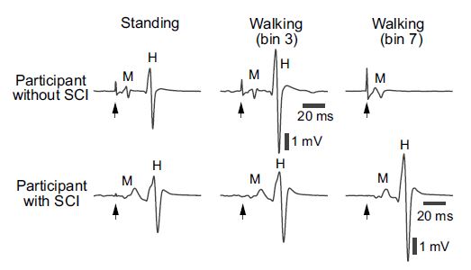
Figure 2 of the paper showing examples of the soleus H-reflex (labeled H) during standing and during different phases of walking in a participant without known neurological injuries (top) and in a participant with chronic incomplete SCI (bottom).
Today is my third attempt at a critical review paper. Since my PI gets a copy, so do all of you! You can read my first looking at elbow spinal stretch reflexes here. Or my second where I review modulating spinal cord excitability with a static magnetic field here. Today is an interesting paper on soleus stretch reflex and H-reflex. I really appreciate the methodology the researchers used and they did an excellent job of highlighting the limitations to the study, which is always important. Per the usual disclaimer, this is my third critical review, so you can take my opinion n the methodology and findings how you will.
Spasticity, or uncontrolled muscle stiffness is a common issue for people with incomplete spinal cord injury (SCI). This has led to the belief that abnormal reflex activity impairs gait. However, that theory has not been tested. This study looked at modulation of stretch reflex excitability across the entire gait cycle in nine neurologically intact participants and nine with spasticity due to chronic incomplete SCI (ranging from 2.5 – 11 years post injury) classified as D using the American Spinal Injury Association scale. The researchers used electromyography (EMG) to measure stretch reflex while standing and while having participants walk at their own pace on a treadmill. In both conditions, researchers caused an unexpected dorsiflexion perturbation of the ankle at every 4 – 6 steps at different phases of the gait cycle. Soleus H-reflex was also measured using EMG and done by stimulation of the tibial nerve just above M-wave threshold during standing and at different phases of the gait cycle while walking on a treadmill.
Unlike some previous research, they found no statistically significant changes in the H-reflex and stretch reflex in the two populations. However spinal short and medium latency “M1” and “M2” components in the soleus stretch reflexes were found to be abnormally large in the mid-late swing phase in the SCI population. This finding implies that multiple spinal and supraspinal pathways contribute other CNS populations’ spastic movement disorders and could also contribute to the abnormal locomotor reflex modulation the researchers observed. They also suggest that the findings support the possibility that hyperactive spinal stretch reflexes in the mid-late swing phase can impact the following stance phase in the gait cycle.
This study is unique because it is one of the first to look at how spinal stretch reflexes are moderated by dynamic motion (walking) and not just a passive state (standing). The study also highlights that reflexes elicited in a passive state do not necessarily provide information on the function or dysfunction of reflexes during specific phases of dynamic motion. By further understanding how spinal reflexes are modulated, we can better understand the underlying dynamics of other neurological conditions such as clonus.
There were several limitations to the study that the authors acknowledged. The largest was that the data was normalized to the soleus Mmax for standing and not during walking. This was in part due to the limitations of determining Mmax while walking, which would have required significant additional stimuli and steps, thus resulting in a higher risk of fatigue during the experiment for the SCI group. Second, most of the SCI participants were on a stable dose of baclofen and/or other anti-spasticity medications. These medications cause changes to spinal reflex characteristics, which more than likely influenced the findings in this study.
Overall the experimental design was well thought out. The limitations of the data they collected and its analysis, were explained clearly. However, while the main goal of the study was to look at the soleus stretch reflex and H-reflex, by adding several more EMG sensors the researchers could have simultaneously collected data from other large muscle groups, such as: the vastus lateralis, biceps femoris, and gastrocnemius. All of which could have been used to compare the response of the entire leg to the perturbation and not just the change in reflex response of the soleus. This extra information could have been used to determine which muscles modulate the soleus stretch reflex and H-reflex pathways without dramatically increasing the experiment time. This data and analysis could have helped better explain their findings and aid in the conclusions they drew.
Source:
Thompson, A.K., Mrachacz-Kersting, N., Sinkjær, T. et al. Modulation of soleus stretch reflexes during walking in people with chronic incomplete spinal cord injury. Exp Brain Res 237, 2461–2479 (2019). DOI: 10.1007/s00221-019-05603-1


But enough about us, what about you?