Know your spinal cord – Compound action potentials
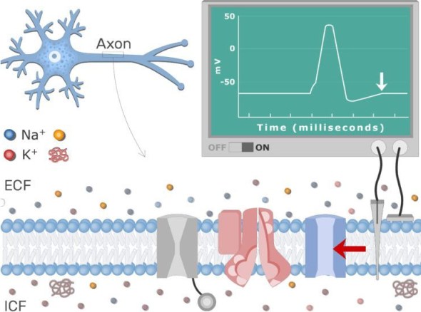
Image from: Get body smart
Day forty-five of the know your spinal cord series is here! With so many posts, you may be wondering how to find them all. Fear not, we have a super helpful neuroanatomy category for all your spinal cord needs. For the past few posts we’ve looked at some very interesting tools to probe the spinal cord. We’ve seen that there are quite a few ways we can go about it, but more importantly they all tell us something slightly different. Today we are looking at the product of that stimulation, the compound action potential.
If you’ve taken an intro to biology course, you may already understand action potentials. While we’ve talked about it as needed, we’ve never really dedicated a whole post on them. That’s what we are doing today. Now there is a difference between an action potential and a compound action potential, but the name somewhat gives it away. A compound action potential is the summation of action potentials generated by a stimulus. As we’ve mentioned, this is a one-way thing, it always travels the same direction for every neuron in all of your body. With one exception, the F-wave, but since that doesn’t occur in nature we can ignore it.
Let’s define action potential a little bit better first. Wikipedia, the source of all human knowledge, defines action potential as “occurring when the membrane potential of a specific cell location rapidly rises and falls: this depolarization then causes adjacent locations to similarly depolarize.” This is a fancy way of saying there is a rapid decrease in electric potential in a cell, which causes the neighboring cell to do the same, creating a sort of negative wave that travels the length of the axon. If a picture is worth a thousand words, then a gif is worth 10,000. Here we see how the action potential travels the length of the axon.

Action potential traveling the length of the axon of a nerve
Let’s talk about how we create this action potential. At rest, the cells in a nerve have what is called a membrane potential . That just means there is a difference between the voltage outside of the cell and the inside of the cell. Below is the wave created by an action potential. The membrane potential (again the difference between the inside of the cell and outside) sits at around -70 mV (1).
If we apply an electrical stimulus we can cause the nerve to fire if and only if it reaches above threshold (2). If we do not rise above threshold, the cell does not fire and we do not create an action potential (not shown below). However, if it does reach threshold or higher we get (3) a rapid rise in voltage.
This follows with an equally quick drop in voltage (4). Then the cell membrane potential drops below what it normally is at rest entering what is called a refractory period (not labeled as such, but 5). While in this period, the cell will not easily fire again because as you can see the voltage required to exceed threshold is much higher while in this stage. Eventually, (after a few hundred milliseconds) the cell returns to a resting membrane potential (6).
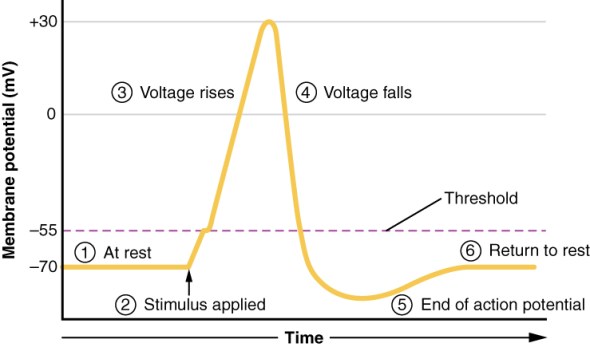
This is what we mean when we say we lower the threshold for firing when doing spinal cord stimulation. Technically we increase the membrane potential of the cells, thus requiring a lower stimuli to create an action potential. This is the basis for nerve communication!
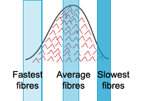
Now that we have the basics of an action potential down, we can talk about the compound action potential! When we are talking action potential we are talking about a single neuron. However, neurons don’t exist in a vacuum. For example, in the spinal cord there are whole pools of neurons that activate when you move. When we record activity in the muscles, from the brain, or even from the spinal cord, unless we are recording using a specific very invasive tool, we are recording the summation of firing across a lot of neurons. This is the basis for the compound action potential. Below is a crude drawing of what this means, basically what we are recording is a lot of single action potentials that create one large envelope response.
Compound action potentials are typically going to be from surface recordings. EMG, EEG, etc. are compound action potentials. We have tools (literally a sharp rod that stabs the cell) to record from a single cell even in vivo, but that is an invasive method and my lab does not use invasive techniques.
As you might imagine, while the image above shows a very smooth curve, compound action potentials (CAP’s) aren’t always this smooth, in fact most of the time they are very complex shapes becuase we may have a lot of fast fibers and slow fibers, but very few average fibers. Below we see the compound action potentials recorded from a muscle (compound muscle action potentials). The shape of this response is much more dynamic then the simplified image above.
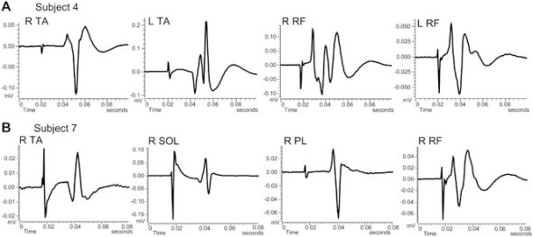
Compound muscle action potentials (CMAPs) evoked by tsMSS. A: Non-rectified waveform averages (n = 10, elicited every 5 s) of CMAPs recorded from the right (R) and left (L) tibialis anterior (TA) and rectus femoris (RF) muscles in subject 4 following tsMSS. B: Non-rectified waveform averages (n = 10, elicited every 5 s) of CMAPs recorded from the R TA, soleus (SOL), peroneus longus (PL), and RF muscles in subject 7 following tsMSS. Note the different shape of these compound action potentials across muscles. Time at 0.02 s on the x-axis corresponds to the onset of tsMSS and associated spike of the stimulus artifact. DOI: 10.1002/bem.21768
So now we have a whole post dedicated to what we are recording when we stimulate nerves and what that tells us about the pools of neurons we are stimulating. Overall it is a very complex subject and we’ve just dipped a toe in so to speak. However, the takeaway is that cells need to rise above a certain threshold to fire; after firing for some time, the membrane potential drops even lower meaning it takes more stimulus to get it to fire again; TSS increases membrane potential meaning it takes less stimulus to make the cell fire.
Until next time, don’t stop learning!


But enough about us, what about you?