Know your spinal cord – The spinocerebellar tract
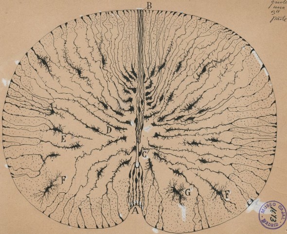
Drawing of a spinal cord cross section by Ramon y Cajal, a famous Spanish neuroscientist.
Here we are on day ten of knowing your spinal cord. As always, you can find the aggregated posts in my totally new, fresh off the line category, neuroanatomy. There posts are organized in reverse chronological order, so the first post on the medullary pyramids would be at the bottom, which is where I recommend you start if you’re new. For everyone who’s followed along or those of you who are just interested in this one tract, let’s talk about the spinocerebellar tract!
Not to be confused with another spino- tract we’ve already covered (the spinothalamic tract) this is a whole different tract in the spinal cord. This tract, like the spinothalamic tract is an ascending or afferent pathway. If we knew nothing about this tract, we could tell from the name, based on the way it is named we know where it starts, spino- involving the spine, and where it ends -cerebellar (spoiler alert), involving the cerebellum.
Even though we skipped over this tract a few posts back when we covered the white matter it is actually a complex tract that is normally subdivided into four parts, the dorsal, ventral, Cuneo-, and Rostral spinocerebellar tracts. Each of these four subdivisions handle something slightly different so today we will take them one at a time.
First let’s talk about the naming convention. You can tell a lot about a particular tract based on how it is named. Like I’ve just shown the name of the tract can tell you where it originates and where it terminates. It can also tell you where it is located, when we talk about location we use fancy terms, but once you remember them they are easier to use than some other naming convention (left and right for example) because of how we define them.
Dorsal, this is the same thing as saying towards the rear and sometimes is interchanged with posterior because posterior means roughly the same thing. Ventral is the opposite direction to dorsal, meaning towards the front and is sometimes interchanged with anterior for the same reason dorsal and posterior are sometimes interchanged. Rostral is how we define one of the other axis, this means toward the head (if we are in the brain, this would mean towards the front of the brain). If we want to go in the other direction, we say that is caudle. Below we have an image showing these different directions.

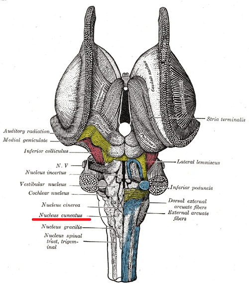
This leaves one pathway, the Cuneocerebellar pathway without a direction, that is because the term Cuneo- isn’t in reference to a direction, but to a location! This is in reference to the accessory cuneate nucleus, which is found in the medulla lateral to the cuneate nucleus. Cuneo- meaning wedge, which refers to the shape of the nucleus. You can see the location of the cuneate nucleus below, which I’ve underlined in red to make it easier to find.
So now that we have a good idea about where the tracts are located (based on the name), let’s take a look at them one at a time and talk about what they do. Below is an image showing a cross section of the cord. The tract has only been subdivided into two of the four divisions we’re talking about today, but they are in the same portion of the spinal cord.

Dorsal spinocerebellar tract
The dorsal spinocerebellar tract runs parallel with the ventral spinocerebellar tract. It innervates two different types of cells that deal with proprioception they are muscle spindles and Golgi tendon organs. As a refresher proprioception is how we tell where are limbs are in space and is essentially done by measuring stretch of the muscle.
To that end, muscle spindles are literally defined as stretch receptors in the muscle that detect changes in the length of the muscle. These signals are sent through type 1a afferent sensory fibers (image below) and are partially responsible for reflexes, which we will cover in detail in another post.

However, there is another proprioceptive sensory organ involved called the Golgi tendon organ. This guy does things slightly different and instead of dealing with stretch, he measures tension in the muscle. Like the type 1a afferents, the Golgi tendon organ has its own reflex aptly named the Golgi tendon reflex mediated by type 1b afferents (which is why we have a and b). They look quite a bit different to the muscle spindles, since they weave into the collagen cells, which looks sort of like a woven basket. We can see that in the image below.

So how does this tract run? First proprioceptive information is passed to the spinal cord through the dorsal root ganglia (this would be our first order neuron). They travel through the dorsal horn where they synapse with Clarke’s nucleus (here is our second order neuron). Axon fibers from Clarke’s nucleus ( located in lamina VII and found only at the level of T1-L2 on the spinal cord suggesting it only deals with lower limbs) convey this proprioceptive information in the spinal cord in the peripheral region of the funiculus posterior ipsilaterally (same side). The fibers continue to course through the medulla oblongata of the brainstem, at which point they pass through the inferior cerebellar peduncle and finally into the cerebellum, where unconscious proprioceptive information is processed. Notice that this particular pathway is a two neuron pathway, but more importantly it does not decussate (shock!).
Ventral spinocerebellar tract
Now for the ventral spinocerebellar tract. This tract does something similar to the previous one, it conveys proprioceptive information to the cerebellum, so what’s the difference? First, this tract only deals with information from the Golgi tendon organ and has nothing to do with the muscle spindles. It is also a two neuron pathway, however this path isn’t as straightforward as the last tract. The ventral spinocerebellar tract does actually cross to the opposite side of the body first in the spinal cord as part of the anterior white commissure and then cross again to end in the cerebellum (we call this a “double cross”).
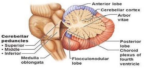
This tract starts similarly, but again only with the Golgi tendon organs. Once it passes through the dorsal root ganglion, it runs via the fila radicularia to the dorsal horn of the grey matter. There it synapses with the dendrites of not one, but two neurons. They send their axons bilaterally to the ventral border of the lateral funiculi. The ventral spinocerebellar tract then enters the cerebellum via the superior cerebellar peduncle, a large bundle of neurons that looks like a stalk, which is where it gets its name (shown below).
Cuneocerebellar tract
Now let’s talk about the cuneocerebellar tract, this portion of the tract deals with proprioceptive information from the upper limbs and neck. Inputs come from both muscle spindles and the Golgi tendon organs. This tract is an analogue to the dorsal spinocerebellar tract, but for the upper limbs. If you notice, we mentioned earlier that the dorsal tract synapse on Clark’s nucleus, which is only located in the level of T1-L2 on the spinal cord. This pathway is also a two neuron pathway and synapses at the accessory cuneate nucleus (shown above underlined in red close to the beginning of this post). This means it does not synapse in the spinal cord like most of the tracts we’ve covered. However, like its sister the dorsal spinocerebellar tract, the cuneocerebellar tract is runs ipsilateral!
Rostral spinocerebellar tract
Now for the last tract, the rostral spinocerebellar tract. We’ve already covered two tracts that use both muscle spindles and Golgi tendon organs for sensory input. However, we’ve only covered one (the ventral spinocerebellar tract) that has input from the Golgi tendon organs exclusively, this tract is the sister tract for the ventral spinocerebellar tract in that it only uses Golgi tendon organs for sensory input. The last two tracts have dealt with upper limb and lower limb exclusively. This tract is similar, it relays proprioceptive input primarily from the ipsilateral head and upper limb as well as movement of head and upper limb (again ipsilateral or same side).
How does this tract travel? Proprioceptive information from the head and upper
limb is transmitted by the Golgi tendon organs to C4–C8 spinal cord levels. This is our first order neuron and the cell body is in the dorsal root ganglion, these synapse with second order neurons whose cell bodies reside in lamina VII of the dorsal horn. The fibers of the second order neurons make up the ipsilateral rostral spinocerebellar tract, which as we mentioned is the head and upper limb counterpart of the ventral spinocerebellar tract. These fibers join at the inferior cerebellar peduncle and synapse at the cerebellum.
Told you this was going to be a complex tract! Now for the takeaway, if you remember anything from this post, just remember this. Most of the proprioceptive information does not reach conscious levels. Instead the information is transmitted directly to the cerebellum via the ascending somatosensory cerebellar pathways without projecting to the thalamus or the cerebral cortex. Lastly, these pathways, which process subconscious proprioception from muscles, tendons, and joints, are only thought to be two-neuron pathways.
Next up I think we will cover spinal reflexes, but that could change. I’m not sure yet. You’ll just have to wait and see.
Until next time, don’t stop learning!


But enough about us, what about you?