Know your spinal cord – The reflex pathways
Beautiful spinal cord art, used with permission by Greg Dunn design.
Day eleven, we’ve almost spent two weeks covering your spinal cord! Tomorrow we will take a brief break as every two weeks I have a review paper due to my PI in the spinal cord feild, so tomorrow I will share it with all of you as well. For today, remember we have a neuroanatomy category with all of the posts we’ve done. If you’re new I would suggest you start with the medullary pyramids post and work forward, for the rest of you or those of you who are only interested in reflexes, let’s talk about some legos.
Was that a typo? No, we’re really talking legos for the moment. I want to highlight something you may not have realized. If you’ve ever stepped on a lego (or anything particularly painful) you know that you immediately withdraw your foot. What you may not realize is that you do this before you are consciously aware you stepped on something. That is because nerves are rather slow. In fact we’ve measured this and we know for a fact that you cannot consciously respond (IE human reaction time) to a stimulus in under 100 ms.
Now 100 ms might not seem that long, because frankly it isn’t for us. However, it is forever in the terms of your brain. If you touched something very hot, 100 ms before you can react is forever and can cause serious damage to the body. Thankfully we don’t have to pass all information off to the brain to make decisions. Reflex responses are handled in the spinal cord, making them much, much faster.
There are several different types of reflexes, not just a withdrawal response we demonstrated. You may have had your knee hit by the doctor and watched as your leg jumped all on its own. That is a different type of reflex called the stretch reflex. These reflexes are one way we study how the spinal cord works and we’ve even covered a recent paper that studied a stretch reflex pathway. Now that we’ve introduced them, let’s look at a few of the pathways and how they work so quickly!
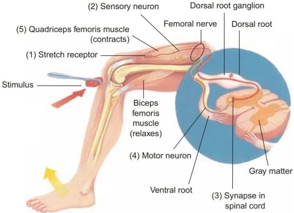
Reflexes can be broken down broadly into two categories, monosynaptic and polysynaptic. Monosynaptic reflexes (also called deep tendon reflexes) are defined as consisting of only one sensory neuron and one motor neuron, so by activating the sensory neuron, the motor neuron responds. We just gave an example of this, the patellar reflex is a monosynaptic reflex. Brief stimulation to the muscle spindle (a tap to the tendon for example) results in a contraction of the agonist or effector muscle. Below we show that pathway, notice how it is basically just a loop, monosynaptic reflexes are thought to be very simple reflexes (although that is debatable in my opinion).
Do not confuse this with the golgi tendon reflex, which is an inhibitory reflex. The golgi tendon organs measure tension and this reflex causes relaxation of a muscle before the tendon tension becomes high enough to cause damage. It is less sensitive than the stretch reflex, but it can also override the stretch reflex. This is part of the reason why you cannot flex opposing muscle groups.
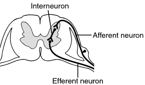
In contrast, more “complex” reflexes are polysynaptic. Polysynaptic reflex pathways involve one or more interneurons that connect to afferent (or sensory) and efferent (motor) signals. Almost all reflexes are polysynaptic. For those who don’t recall, interneurons are neurons that are in the middle (hence inter-) of other neurons. Below is an example of what this looks like.
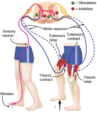
For example, our lego reflex (okay, technically it’s called the withdrawal reflex, but you get it) is polysynaptic. When you step on something (or touch something) that causes pain, the pain receptors in the skin trigger an impulse that travels to the spinal cord. The nerves synapse with interneurons, these neurons are connected to motor neurons. Some of these send motor impulses to the flexors that lead to the muscles in the leg to contract (or withdrawal from the noxious stimulus). However, that’s not the end of it, other motor neurons send inhibitory impulses to the extensors so that flexion is not inhibited. This is referred to as reciprocal innervation. Below we lay out the pathway for the leg.

Moreover, the strength of the withdrawal reflex is correlated with the strength of the pain. This is one reason I like to use the lego example, becuase let’s face it is there a greater pain? However, you just pulled your leg away, but somehow you don’t fall over, why?
The answer is, of course, another reflex! This one is called the crossed extensor reflex. Let’s start from the top, once you activate the withdrawal reflex, you also activate this reflex. Let’s say that pain receptors are activated (also called nociceptor), the signal travels to the dorsal horn, like we have seen in the past. The nerve synapses with the ipsilateral motor neurons that exit the ventral horn, this is the withdrawal reflex and it happens in 50 ms (or less!).
So we’ve just synapsed on this ipsilateral motor neuron, the same nerve (the one that synapses on the ipsilateral side) also synapses on a motor neuron in the contralateral (opposite side) anterior horn. This motor neuron is in control of the uninjured side of the body and helps stabilize it. In other words, you withdrawal from stepping on the lego and your other leg contracts reflexively to support the weight of your body.
But wait there’s more! At the same time as these two motor neurons are activated, the sensory neuron also sends signal along the cord to other motor neurons to contract muscles that help shift the center of gravity to help maintain balance. In particular, this contralateral stimulation of motor neurons to stabilize the body is what we call the crossed extension reflex. As you might imagine, this particular reflex is usually limited to the lower extremities. Below shows how this reflex pathway runs.
If this reflex pathway sounds complex, that’s becuase it is! Reflexes are extremely complex and the more we study them the more we realize that they are not as simple as we thought they were. There are also other reflexes that we are born with and lose as we become adults (like the grasping reflex babies have). If you’ve ever taken martial arts, any sort of sport, or even music, you know that you can even learn reflexes! To me this speaks to just how amazing the spinal cord is becuase it gives us a glimpse into what we’ve just recently realized in the past five years or so. The spinal cord not only sends, receives, processes, and responds to sensory and motor commands, it also is capable of learning independently of the brain.
We can talk about spinal learning next time, but for now this is just a glimpse into some of the main reflex pathways you may deal with day to day. Just a reminder tomorrow is a new review on a recent(ish) paper on the spinal cord, but after that it’s back to the neuroanatomy. At least until we cover everything I can think to cover, which is probably a few more weeks worth of posts at least.
Until next time, don’t stop learning!


Beautiful! 🙂
LikeLike
February 2, 2020 at 4:18 pm
I’m assuming you mean the first image and I agree. I wish I had artistic talent, but I do not.
LikeLike
February 2, 2020 at 5:13 pm
The first image yes, but also the human body has a beauty all its own. 🙂
LikeLike
February 2, 2020 at 5:16 pm
Agreed! It’s just mind boggling how it all came together the way it did.
LikeLiked by 1 person
February 2, 2020 at 7:17 pm