Know your spinal cord – The meninges
Day sixteen of knowing your spinal cord! So many posts, much knowledge! For those who are just finding us, we have a whole neuroanatomy category dedicated to these posts. If you want to take it from the top (literally) you should start with the medullary pyramids post. If you’ve been following along or only interested in this particular topic today we are covering the meninges and you’ll learn my really dumb way for remembering them, so let’s get started.
The meninges, you don’t really think about them when you’re thinking about the brain or the spinal cord, but they are very important. In fact, these are the things that keep your brain and spinal cord safe, held in place, and separated from the body. Depending on your background you may know there are three meninges that surround the brain and the spinal cord. Most people neglect to mention they are shared between the brain and the spinal cord, but not us! You may be wondering why there are three, well that’s a good question and we can take them one at a time to give you the full picture.
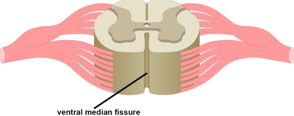
Let’s work inside out, that is how I like to remember them (I will give you that bit of information at the end of the post). So first up let’s talk about the pia mater. Latin for “tender mother” the pia mater is the very delicate innermost layer of the meninges. Originally part of something called the neural crest, a temporary group of cells unique to vertebrates that give rise to a very diverse cell lineage, the pia mater is permeable to both water and small solutes. It closely follows and encloses the curves of the spinal cord, and is attached to it through a connection to the ventral (or anterior) median fissure. Below is an image showing the ventral median fissure for those of you who need a refresher.
The pia mater of the spinal cord is slightly different than the brain. I won’t go into details of how they are different, suffice to say they are still very similar. The pia mater doesn’t actually attach to the next layer (the arachnoid mater). Instead it attaches to the outer layer, the dura mater through 21 pairs of denticulate ligaments, which are bilateral (both sides) triangular extensions of pia mater. Below is an image showing the denticulate ligaments. The denticulate ligaments help to anchor the spinal cord and prevent side to side movement, which provides stability, a common theme of the meninges.

The membrane of the spinal cord is much thicker than the cranial pia mater, due to the two-layer composition of the pia membrane. The outer layer, made up of mostly connective tissue, is responsible for this thickness. Between the two layers are spaces which contain the cerebrospinal fluid (CSF) and is called the subarachnoid cavity. This is just the space between the pia and arachnoid mater.
At the point where the pia mater reaches the conus medullaris (the end of the spinal cord), the membrane extends as a thin filament, which is called the filum terminale or terminal filum, contained within the lumbar cistern. This filament eventually blends with the dura mater and extends as far as the coccyx (tailbone). It then fuses with the periosteum, which is a membrane found at the surface of all bones, and forms the coccygeal ligament. There it is called the central ligament and assists with movements of the trunk of the body. You can see what this looks like below.

Lastly we should mention that the pia mater also functions to deal with the deformation of the spinal cord under compression. Due to the high elastic modulus of the pia mater, it is able to provide a constraint on the surface of the spinal cord. This constraint stops the elongation of the spinal cord.
Now let’s talk about the arachnoid mater. This is also a derivative of the neural crest (hint: the dura mater is not, which is why I bring it up). If the name doesn’t give it away, it is named for the way it looks. Once again scientists aren’t creative when it comes to names and this is a good thing. The arachnoid mater has a spider web-like appearance, which is easy to remember because of the name. It is a thin, transparent membrane is composed of fibrous tissue and, like the pia mater, is covered by flat cells also thought to be impermeable to fluid. It mainly serves as a cushion for the brain and spinal cord, but it has other purposes as well.
Cerebrospinal fluid circulates in the subarachnoid space (between arachnoid and pia mater). We didn’t cover this yesterday, CSF but is produced by the choroid plexus (inside the ventricles of the brain), which are in direct communication with the subarachnoid space so the CSF can flow freely through the nervous system. Unlike the pia mater, the arachnoid mater doesn’t sit as closely to the surface it covers, so it looks more like a sac. The arachnoid mater and dura mater are very close together all the way to around S2 of the spinal cord, where the two layers fuse into one and end in the filum terminale.
This brings us to the dura mater. Latin for “tough mother,” (don’t ask why they got stuck on the mother naming) it is the outermost layer of the meninges. As its name suggests, it really it tough, in fact out of the three layers it is the strongest. It is particularly thick when compared to its two counterparts and also sits very loosely around the brain and spinal cord. Unlike the other two layers, this one is derived from the embryonic mesoderm (one of the three primary germ layers in the very early embryo)!
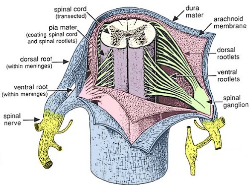
Technically when we are referring to the spinal cord, it is called the dural sac and is comprised of two layers. The inner layer is the meningeal layer and the outer one is called the periosteal layer. The space between these two layers is called the epidural space. This is where the main differences between the cranial meninges and the spinal meninges come into play. Unlike the cranial epidural space, the spinal epidural space contains adipose tissue (fat) and the internal vertebral venous plexuses (intraspinal veins). Below we can see all three of the spinal meninges labeled. I like this image a lot becuase you can see that the meninges also cover the dorsal and ventral roots.
Now that we’ve introduced you to the three meninges and how they function, I have a video of the dissection of an unfixed spinal cord. Another video by Dr. Stensaas, which I cannot recommend her video lectures enough. It shows an actual spinal cord, goes into detail about the meninges and gives you a great overview. Plus, a lot of us like to think that the spinal cord is thicker than it actually is, it is really about the size of your pinky (as she demonstrates), but the video does a great job of showing just how thin and delicate the spinal cord really is.
That about wraps it up for today. Sticking with the theme lately, I’m not sure what I will be talking about tomorrow, but I’ll figure it out. In the meantime, I should give you the easy way I like to remember the order of your meninges. It’s stupid, so you will never really forget it, but P.A.D. This tells us all about the meninges, they act primarily as padding and going from the inner to the outer we have the Pia, Arachnoid, and Dura mater, or P.A.D. Now that I’ve given away my secret, you’ll always remember the correct order and the primary function all in one acronym.
Until next time, don’t stop learning!


But enough about us, what about you?